
Spot Test Through Microscopic Slides: Most Common Slides in Microbiology and Their Images with Main Features

Spot Test Through Microscopic Slides
‘Spot Test Through Microscopic Slides’ is the most common microscopic slide in Microbiology and its images with main features. This spot test is very useful for both bachelor as well as master degree programs i.e. B.Sc. MLT/ BMLT or MBBS or M.Sc. MLT/ M.Sc. Clinical/ Medical Microbiology, etc. This collection of spots makes your examination easy.
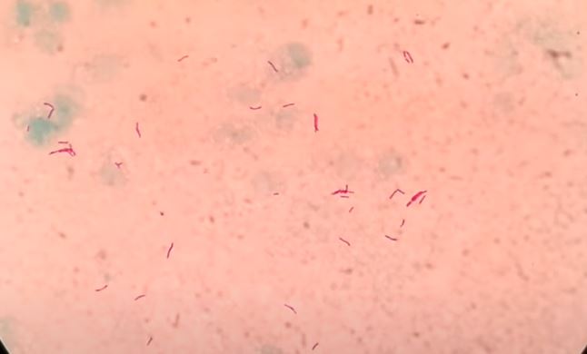
Spot Test: 1

Salient features
- Violet in color
- Cocci are in singles, pairs, chains, and clusters.
- The most probable organism is Staphylococcus aureus while Streptococcus pyogenes (in chains ), and Streptococcus pneumonie (in pairs).
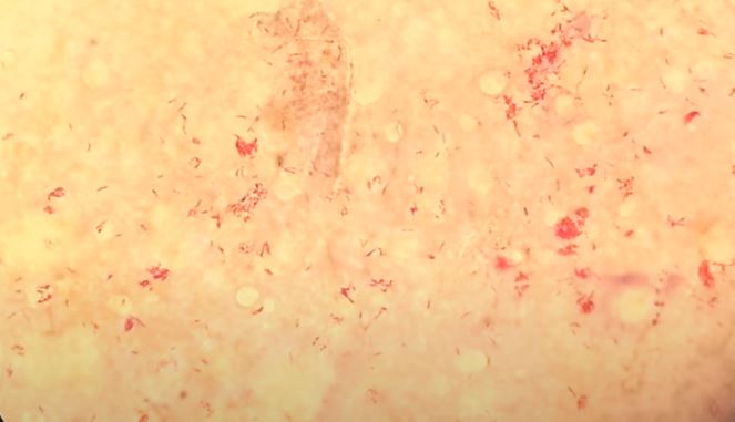
Spot Test: 2

Salient features
- Red in color and rod in shape.
- So organisms are Gram-negative bacilli.
- Bacteria may be E. coli, Klebsiella, Proteus etc
Spot Test: 3

Salient features
- Red in color and cocci in shape, arrangement in pairs.
- Present intracellularly as well as some diplococci are also present extracellularly.
- Possible organisms may be Neisseria gonorrhoeae and Neisseria meningitides.
Spot Test:4
Main Features
- Acid-fast bacilli (AFB), Pink, beaded, thin slender rod with some are curved having size about 1 -8 x 0 .2 -0 .6µm.
- The causative agent of tuberculosis
- The common culture medium is the LJ medium.
- It takes 3-6 weeks to grow on LJ medium.
Spot Test:5
Principal Features
- Acid-fast bacilli (AFB), more than 1000 bacilli, clumps, and globi in every field
- 1% acid alcohol or 5% sulphuric acid is used for Z-N staining.
- It is the causative agent of leprosy.
- It can not grow on inanimate media.

Salient Features
- Albert’s staining is used to identify Corynebacterium diphtheriae.
- The causative agent of diphtheria.
- Selective medium: Potassium tellurite blood agar.
- Another medium used: Loeffler serum slope (LSS).

Salient features
- Gram-positive yeast cells.
- Having the size of nearly 3-8 µm.
- Opportunistic fungus.
- The causative agent for oral thrush and vaginitis.

Salient features
- Germ tube test(GTT)is used to identify mainly Candida albicans.
- Candida tropicalis may produce pseudo-germ tubes after 3 hrs of incubation but they show constriction at the point of origin.
- Another germ tube test positive Candida species is C. dubliensis but it can be differentiated by culturing at 42°C where C. dubliensis can not grow but C. albicans grow.
- This test is also named as Reynold- Braude phenomenon.
Salient features
- Cryptococcus neoformans is an encapsulated yeast.
- The causative agent of meningitis called cryptococcal meningitis.
- It is Gram-positive.
- It can easily grow on Sabouraud’s dextrose agar (SDA).
- Produces disease in mice (animal inoculation test positive).

Main features
- Female gametocyte, also called macrogametocyte is banana or sickle-shaped, longer, and more slender.
- The cytoplasm stains deep blue and the nucleus is compact.
- The female anopheles mosquito is the definite host whereas the man is the intermediate host.
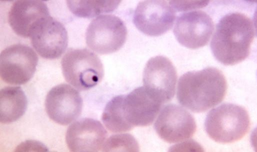
Principal Features
- Cytoplasm opposite the nucleus is thicker in early trophozoite or ring form.
- Young RBCs are infected and these are enlarged.
- It causes malaria.
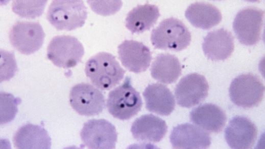
Main Features
- Multiple rings in a single RBC.
- No age matter of RBCs i.e. any age of RBCs get infected.
- The invaded cell is not enlarged.
- It can cause cerebral malaria, blackwater fever and is also responsible for most cases of resistant malaria.
Salient Features
- Leishmania donovani bodies (LD bodies) are flagellar (Amastigote ) stage of Leishmania donovani.
- It is a round or oval body ( 2 to 4 μm).
- Present in the cells of the reticuloendothelial system ( liver, spleen, and bone marrow) of a man suffering from Kala-azar or Visceral leishmaniasis.
- The insect vector for Kala-azar is sandfly.

Salient Features
- Microfilaria of Wuchereria bancrofti is an embryo of Wuchereria bancrofti.
- It measures 290 μm × 6-7 μm in size.
- Covered by a hyaline sheath which is much longer than the embryo.
- The somatic cells or nuclei extend from the head to the tail end.
- The nuclei do not extend up to the tip of the tail, a distinguishing feature of Microfilaria of Wuchereria bancrofti.
Rhinosporidiosis: sporangium with numerous endospores
Salient features
- Causative agent is Rhinosporidium seeberi.
- Causes rhinosporidiosis, a chronic granulomatous disease characterized by friable polyps, usually confined to the nose, mouth or eye.
- The majority of infections occur in persons who have frequent contact with stagnant water or aquatic life.
Contd…
[5233 visitors]