
Nigrosin Preparation Showing Cryptococcus Capsule: Introduction, Preparation and Result Interpretation
Cryptococcus capsule in Nigrosin
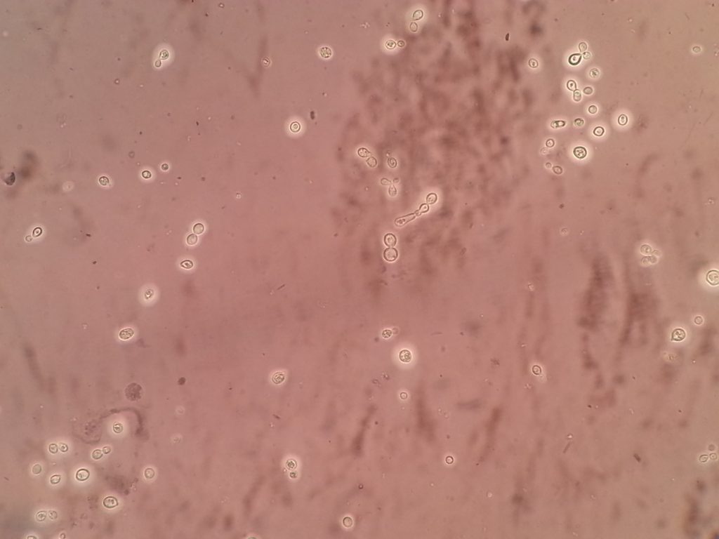
Nigrosin preparation-Positive with the following characteristics-
Distinct, wide gelatinous capsules
Clinical specimen-Cerebrospinal Fluid (CSF)
India ink or nigrosin preparation (negative staining) for Fungal Infections Laboratory Diagnosis
India ink or nigrosin preparation may be used to examine CSF for the presence of the encapsulated yeast such as Cryptococcus neoformans. With negative stain, budding yeast surrounded by a large clear area against a black background is presumptive evidence of C. neoformans as shown here.
Test Procedure
In CSF for Cryptococcus neoformans:
- First centrifuge the CSF.
- Take a drop of sediment part on a clean and grease-free slide and add over it a drop of nigrosin.
- Mix it properly and put a coverslip over it.
- Observe under a microscope, focusing at 10X objective and finally 40X objective.
In the case of bacteria capsule
Take a clean and grease-free slide. Put a drop of nigrosin over it. Touch the colony with an inoculating loop from a solid medium or take a loopful broth in case of a liquid medium. Mix the nigrosin drop and make a smear. Leave for air dry. Focus at 10X objective and finally observe at oi immersion lens i.e. 100X objective.
Principle And Interpretation
Negative staining permits visualization of the usually transparent and unstainable capsule of many organisms, most importantly Cryptococcus neoformans. Nigrosin consists of a suspension of fine particles of carbon. These form a dark background, against which capsules see clearly as a result of displacement of the carbon particles.
Quality Control
Appearance blackish violet-colored solution.
Clarity
Clear without any particles.
Results
It is clear halos surrounding the cells as shown above image.
Further Readings
- Medical Mycology. Editors: Emmons and Binford, 2nd ed 1970, Publisher Lea and Febiger, Philadelphia.
- Rippon’s JW: Medical Microbiology. The pathogenic fungi and the Pathogenic Actinomycetes. 3rd ed 1988 Publisher WB Saunder co, Philadelphia.
- Clinical Microbiology Procedure Handbook Vol. I & II, Chief in editor H.D. Isenberg, Albert Einstein College of Medicine, New York, Publisher ASM (American Society for Microbiology), Washington DC.
- A Textbook of Medical Mycology. Editor: Jagdish Chander. Publication Mehata, India.
- Practical Laboratory Mycology. Editors: Koneman E.W. and G.D. Roberts, 3rd ed 1985, Publisher Williams and Wilkins, Baltimore.
- Topley & Wilsons Medical Mycology. Editors: M.T. Parker & L.H. Collier, 8th ed 1990, Publisher Edward Arnold publication, London.
- Textbook of Diagnostic Microbiology. Editors: Connie R. Mahon, Donald G. Lehman & George Manuselis, 3rd edition2007, Publisher Elsevier.
- Mackie and Mc Cartney Practical Medical Microbiology. Editors: J.G. Colle, A.G. Fraser, B.P. Marmion, A. Simmous, 4th ed, Publisher Churchill Living Stone, New York, Melborne, Sans Franscisco 1996.
- Bailey & Scott’s Diagnostic Microbiology. Editors: Bettey A. Forbes, Daniel F. Sahm & Alice S. Weissfeld, 12th ed 2007, Publisher Elsevier.