
Candida albicans: Introduction, Morphology, Pathogencity, Laboratory Diagnosis and Treatment
Introduction of Candida
The yeast is a common commensal of the gastrointestinal tract. Most Candida species are opportunistically occurring in debilitated persons e.g. Diabetes patients, those who are taking anticancer therapy and immunocompromised patients, HIV patients, and so on.
Classification of Candida
Scientific classification
Kingdom: Fungi
Division: Ascomycota
Class: Saccharomycetes
Order: Saccharomycetales
Family: Saccharomycetaceae
Genus: Candida
Species: C. albicans
Other medically important Candida species are Candida albicans, Candida tropicalis, Candida parapsilosis, Candida glabrata and Candida krusei.
Morphology of Candida
Yeasts are small, oval, measuring 3-4 µm in diameter. Single, budding of the cells may be seen. The yeast cells can also be seen attached with pseudohyphae.
Pathogenicity of Candida albicans
It causes a disease called candidiasis also called moniliasis. It is an infection causing fungi of the genus formerly Monilia or now Candida (especially Candida albicans).
The various forms of diseases are –
- Oral thrush: Also known as Candidiasis of the mouth or oropharyngeal candidiasis which is seen as white patches on the mucosa of the mouth including the tongue. The affected site can become inflamed and may cause difficulty in swallowing causing cracking and inflammation which may occur around the mouth. Such a condition is referred to as oral cheilitis. Oral thrush may spread to the esophagus (esophagitis). Although most people harbor Candida species, oral candidiasis is typically found in immunocompromised hosts like AIDS patients (9 to 31% ), people taking immunosuppressive drugs for cancer chemotherapy (20%), and organ transplantation. Other factors associated with oral thrush are diabetes, certain dentures, and the use of corticosteroids. 5 and 7% of neonates develop oral candidiasis( CDC) and untreated oral thrush can lead to serious invasive disease.
- Vaginal thrush: Also called genital or vulvovaginal candidiasis cause genital itching, a burning sensation, and vaginal discharge in females and while In men, the penis may have an itching rash. This is rare in men but most women will have at least one episode of vulvovaginal candidiasis. Women with the following conditions are more at risk of the infection if they are-√Pregnant
√Diabetic
√Use broad-spectrum antibiotics
√Use corticosteroids - Leucorrhoea: It is a flow of a whitish, yellowish, or greenish discharge from the vagina that may be normal or a sign of infection. Discharges may originate from the various female reproductive parts such as the vagina, ovaries, fallopian tubes, or, most commonly, the cervix. Leukorrhea may occur during pregnancy and is considered normal when the discharge is thin, white, and relatively odorless. Physiologic leukorrhea is a normal condition occurring within several months to a year of the onset of the menstrual cycle in adolescent girls and is sometimes present in newborn girls, usually lasting one to two months. However, in many cases, leukorrhea is a sign of infection, especially when the discharge is yellow or green, with an offensive odor, and is accompanied by irritation, itching, pain, or tissue inflammation due to Candida.
- Candidemia: Also called Invasive candidiasis is a serious disease when Candida, which is normally on the skin or the gastrointestinal tract (GIT), enters the bloodstream where it can disseminate to other organs. Patient with such condition has symptoms like fever and chills that do not respond to antibacterial agents. These are often nosocomial ( hospital-acquired) infections of people who:√have a central venous catheter√are immunosuppressed√take broad-spectrum antibiotics√show neutropenia√are on hemodialysis√have diabetes
- Meningitis and meningoencephalitis: Meningitis due to Candida is mucocutaneous and deeply Invasive Candidiasis are uncommon. Infection can be secondary to hematogenous dissemination or direct inoculation. Neurosurgery, recent antibiotics, and corticosteroids are predisposing factors. Fever, meningismus, elevated CSF pressures, and localizing neurologic signs are commonly noted.
- Vaginitis particularly during pregnancy
Laboratory Diagnosis of Staphylococcus aureus
Sample collection
Samples are collected according to the site of infections. They may be-
- vaginal swab
- Tongue swab
- Blood
- CSF
- Tissue
- Urine
- Exudate
- Swabs from the mucosal surface
Direct microscopic examination
Wet mount preparation
Gram stain
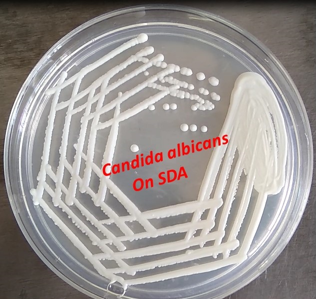
Culture: Culture on Sabouraud Dextrose agar (SDA) at 37° C for 24-48 hours. After incubation observes colonial morphology.
Colony characteristics
Cream-colored pasty and glistening as shown above picture.
Identification of Candida albicans
Wet mount preparation: Single or budding yeast with or without pseudohyphae.
Gram stain: single or budding yeast cells with or without pseudohyphae and gram-positive
Germ tube test: Positive
The test is carried out using 0.5 ml rabbit or human serum in which test yeast cells are inoculated and incubated at 37°C for 2-3 hours.
Put a drop of this after 2-3 hours incubation on the slide and cover with the coverslip. Focus at 10X objective and finally observe at high power objective (40X) of a compound microscope.
Result Interpretation
Germ tube test (GTT) positive: Presence of sprouting yeast cells
Germ tube test negative: Absence of sprouting yeast cells
Chlamydospore formation
It forms in Cornmeal tween agar after incubation for 48-72 hours at 22-25°C. Chlamydospores are spherical, thick-walled, and usually produced on supporting cells that occur along pseudohyphae or at the tip of hyphae.
Treatment
Treatment of Candidiasis depends on location and severity.
For oral thrush–Oral nystatin suspension
Similarly for skin and vulvovaginitis –topical antifungals while in resistant case
azole antifungal medication
In severe infections
- Amphotericin B
- azole antifungals
- Echinocandins like micafungin
Keynotes
- All fungi are gram-positive.
- To diagnose Moniliasis, a serological test in patient serum to detect the antibody to Candida albicans should perform. Four folds rise in titer of antibody in paired sera of the patient is diagnostic.
- CHROMagar Candida or HiCrome candida differential agar recommendation is for rapid isolation and identification of Candida species from mixed cultures in clinical and non-clinical samples.
- For Candida spp. identification other physiological tests like sugar (glucose, galactose, sucrose, maltose, lactose, trehalose) fermentation, and assimilation tests are used.
- Differences between hyphae and pseudohyphae are in a table.

Further Readings
- Medical Mycology. Editors: Emmons and Binford, 2nd ed 1970, Publisher Lea and Febiger, Philadelphia.
- Rippon’s JW: Medical Microbiology. The pathogenic fungi and the Pathogenic Actinomycetes. 3rd ed 1988 Publisher WB Saunder co, Philadelphia.
- Clinical Microbiology Procedure Handbook Vol. I & II, Chief in editor H.D. Isenberg, Albert Einstein College of Medicine, New York, Publisher ASM (American Society for Microbiology), Washington DC.
- A Textbook of Medical Mycology. Editor: Jagdish Chander. Publication Mehata, India.
- Practical Laboratory Mycology. Editors: Koneman E.W. and G.D. Roberts, 3rd ed 1985, Publisher Williams and Wilkins, Baltimore.
- Topley & Wilsons Medical Mycology. Editors: M.T. Parker & L.H. Collier, 8th ed 1990, Publisher Edward Arnold publication, London.
- Textbook of Diagnostic Microbiology. Editors: Connie R. Mahon, Donald G. Lehman & George Manuselis, 3rd edition2007, Publisher Elsevier.
- Mackie and Mc Cartney Practical Medical Microbiology. Editors: J.G. Colle, A.G. Fraser, B.P. Marmion, A. Simmous, 4th ed, Publisher Churchill Living Stone, New York, Melborne, Sans Franscisco 1996.
- Bailey & Scott’s Diagnostic Microbiology. Editors: Bettey A. Forbes, Daniel F. Sahm & Alice S. Weissfeld, 12th ed 2007, Publisher Elsevier.
- https://www.microbiologybook.org/mycology/mycology-3.htm
- https://www.sciencedirect.com/topics/medicine-and-dentistry/candida-meningitis
- https://www.britannica.com/science/leukorrhea