
Saprochaete capitata:Introduction, Morphology, Pathogenicity, Lab Diagnosis, Treatment, Prevention, and Keynotes
Introduction
Saprochaete capitata is an emerging opportunistic fungal pathogen that causes severe, frequently fatal systemic infections in profoundly immunocompromised individuals. It is notorious for its intrinsic resistance to echinocandins and high resistance to fluconazole, which often results in breakthrough fungemia during standard empirical antifungal therapy.
- Synonyms: Formerly known as Geotrichum capitatum, Blastoschizomyces capitatus, and Blastoschizomyces capitis. Its current accepted teleomorph name is Magnusiomyces capitatus.
- Habitat: Ubiquitous in nature, isolated from soil, water, plants, air, and dairy products.
- Human Colonization: Acts as a transient component of the normal human mycobiota, colonizing the skin, gastrointestinal tract, and respiratory tract.
- Clinical Significance: It behaves as an aggressive iatrogenic pathogen in specialized hospital units (such as hematology-oncology wards).
Morphology
Macroscopic Features (Cultural Characteristics)
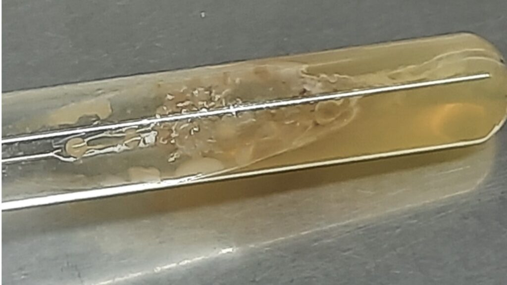
- Sabouraud Dextrose Agar (SDA): Grows moderately fast at 30°C–37°C. Colonies appear cream-colored to whitish, dry, flat, wrinkled, and finely suede-like with irregular or slightly filamentous borders.
- Temperature Tolerance: Uniquely capable of growing at elevated temperatures up to 42°C–45°C.
- CHROMagar: Produces distinct white to cream colonies, aiding in differentiation from other species.
Microscopic Features
- True Hyphae & Pseudohyphae: Profusely branched at acute angles with clear septations.
- Arthroconidia: Abundant, rectangular or cylindrical, single-celled, hyaline thallic conidia formed by the fragmentation of hyphae.
- Blastoconidia / Annelloconidia: Born on long, cicatrized rachids (specialized conidiogenous structures) extending from the corners of arthroconidia or hyphae.
Pathogenicity and Clinical Features
Risk Factors
- Profound Neutropenia: Usually occurring post-chemotherapy with an absolute neutrophil count <500/mm³.
- Hematological Malignancies: Primarily seen in patients with Acute Myeloid Leukemia (AML) and Acute Lymphoblastic Leukemia (ALL).
- Other Factors: Central venous catheters, broad-spectrum antibiotic usage, and breakthrough status during echinocandin (e.g., caspofungin, micafungin) therapy.
Pathogenesis
- Barrier Breakdown: Chemotherapy causes severe mucosal damage (mucositis) in the gut or respiratory tract.
- Passive Invasion: Colonizing yeast cells bypass the compromised epithelial barrier to access the bloodstream.
- Angioinvasion: The fungus actively penetrates blood vessel walls.
- Dissemination: It spreads rapidly to deep organs, causing widespread microabscesses.
Clinical Presentations
- Fungemia & Septic Shock: Clinically indistinguishable from bacterial sepsis or candidemia.
- Disseminated Organ Disease: Deep tissue involvement in up to 80% of cases, frequently targeting the lungs (necrotizing pneumonia), liver, spleen, kidneys, and central nervous system.
Laboratory Diagnosis
- Direct Microscopy: KOH mounts, Gram staining, or Calcofluor white stains show true septate hyphae along with characteristic rectangular arthroconidia.
- Biochemical Profiling: It is non-fermentative and urease negative (distinguishing it from Trichosporon spp., which are urease positive).
- Serological Markers: Generates a positive (1→3)-β-D-glucan test but a negative galactomannan test.
- Mass Spectrometry: MALDI-TOF MS provides rapid, highly accurate species identification, though full tube-based ethanol extraction is often required over direct transfer protocols.
- Molecular Identification: Sequencing of the Internal Transcribed Spacer (ITS) region of ribosomal DNA serves as the definitive reference method.
Treatment
Optimal therapy involves early discontinuation of any ongoing echinocandins combined with aggressive, targeted antifungal administration:
| Antifungal Class | Agent(s) | Susceptibility Profile & Therapeutic Role |
| Polyenes | Liposomal Amphotericin B | First-line choice. Often used as primary monotherapy or paired with flucytosine/voriconazole. |
| Extended-Spectrum Azoles | Voriconazole, Posaconazole | Highly Active. Excellent in vitro profiles; widely used for targeted step-down or combination therapy. |
| First-Generation Azoles | Fluconazole | Highly Resistant. Not recommended for empiric or definitive care due to high MICs. |
| Echinocandins | Caspofungin, Micafungin, Anidulafungin | Intrinsically Resistant. Clinical use is contraindicated. |
Prevention
- Infection Control: Adhere to strict hand hygiene and environmental cleaning protocols since the organism forms stable biofilms on hospital surfaces and plastics.
- High-Risk Environmental Monitoring: Track cases closely during hospital construction or renovation work, which can aerosolize fungal elements.
- Antifungal Stewardship: Restrict routine, unmonitored empirical use of echinocandins in leukemia units to minimize selection pressure for breakthrough Saprochaete infections.
Keynotes
- Saprochaete capitata (Magnusiomyces capitatus) is a rare but high-mortality (>50%) opportunistic pathogen.
- It selectively targets patients with severe, prolonged neutropenia and hematological malignancies.
- The hallmark clinical feature is breakthrough fungemia occurring while a patient is receiving echinocandin therapy.
- It mimics Candida macroscopically but is easily differentiated microscopically by its abundant, rectangular arthroconidia.
- It is urease negative, helping separate it from the morphologically similar genus Trichosporon.
- The drug of choice is Liposomal Amphotericin B or Voriconazole; echinocandins are entirely ineffective.
Further Readings
- https://pmc.ncbi.nlm.nih.gov/articles/PMC5131390/
- https://oamjms.eu/index.php/mjms/article/download/oamjms.2019.385/4423/20936
- https://www.reviberoammicol.com/2013-30/248255.pdf
- https://www.mdpi.com/2076-0817/9/11/922
- https://pmc.ncbi.nlm.nih.gov/articles/PMC12220831/
- https://www.jomos.org/articles/mbcb/pdf/2020/02/mbcb200015.pdf
- https://pmc.ncbi.nlm.nih.gov/articles/PMC12255942/
- https://www.researchgate.net/publication/365852371_A_RARE_FUNGAL_INFECTION_REPORT_OF_17_SAPROCHAETE_CAPITATA_CASES
- https://www.sciencedirect.com/science/article/pii/S1130140613000260
- https://www.nature.com/articles/s41598-026-42967-1
- https://pmc.ncbi.nlm.nih.gov/articles/PMC8846490/
- https://journals.asm.org/doi/10.1128/aac.01834-21
- https://mycology.adelaide.edu.au/fungal-descriptions-and-antifungal-susceptibility/hyphomycetes-conidial-moulds/magnusiomyces
- https://europepmc.org/articles/pmc12255942?pdf=render
- https://www.sciencedirect.com/science/article/pii/S1156523316301846
[23 visitors]