
Immunoglobulin: Introduction, Types, Function, Isotypes, Allotypes, Idiotypes , Polyclonal and Monoclonal antibodies

Introduction of Immunoglobulin
Also called antibody
Immunoglobulin is a glycoprotein that is made in response to an antigen and can recognize and bind to the antigen that caused its production.
Protects us from microbial infection.
Are gamma globulins
Synthesized by plasma cells
Constitute 25-30 % of total serum proteins
Antibodies are present in serum, tissue fluids, and mucosal surfaces and on the surface of B-cells where they act as antigen receptors.
The basic structure of Antibody
Immunoglobulin is composed of 4 polypeptide chains.
2 identical light chains (25 kDa each) and 2 identical heavy chains (50-73 kDa each)
Linked by disulfide bonds
Light chains similar in all immunoglobulins
Light chains occur in 2 varieties:-kappa(k) and lambda( λ )
Kappa chains are more frequently found.
Heavy chains:- gamma, alpha, mu, delta and epsilon.
One Ig contains one type of light chain and one type of heavy chain..(each 2/2)
The variable and constant region
Light and Heavy chains are subdivided into variable and constant regions.
Each heavy and light chain contains amino-terminal in variable region carboxy-terminal in the constant region
The variable region extends from N-terminal and the sequence in this region is highly variable.
The constant region extends from the end of the variable region to C-terminal and the amino acid sequence is relatively constant.
Heavy chains are structurally and antigenically distinct for each class.
L and H chains are linked together by both inter and intrachain S-S bonds.
H and L chain domains:-
Each H and L chain is made up of several small but similar regions called domains.
L- chain:- two-domain (VL and CL)
H-chain:- 4 domains in IgA, IgD, IgG (VH, CH1, CH2, and CH3) while 5 domains in IgM, IgE ( VL, CH1, CH2, CH3, and CH4).
Immunoglobulin fold:-
Folded loop-like structure
Hinge region:-
H-chain of arms extends into the hinge region.
Rich in proline and cystine.
Disulfide bond.
Digestion with proteolytic enzymes
Papain cleavage occurs above the S-S bond of the hinge region.
Produces 3 fragments
2 identical fragments called Fab fragments –antigen-binding activity.
Another fragment called Fc fragment (Fraction crystallizable)
Pepsin digestion
Pepsin cleavage occurs below the S-S bond of the hinge region.
Produce a single fragment composed of two Fab like subunits F(ab)2 binds antigen
Fc fragment is not recovered- digested to small numerous peptides.
Classification of Immunoglobulin
- Based on the structure and antigenic nature of the H chain the immunoglobulin is classified into 5 classes.
- IgG (gamma)
- IgA (alpha)
- IgM (mu)
- IgD (delta)
- IgE (epsilon)
Immunoglobulin G (Ig G)
- A most abundant class of Ig in serum
- Constitutes 80% total immunoglobulin
- Present in blood, plasma, and tissue fluids
- Contains less carbohydrate than other immunoglobulins
- It has a half-life of 23 days: the longest of all of the immunoglobulin isotypes
- Crosses placenta and provide natural immunity to fetus and neonate at birth
- Acts against bacteria and viruses by opsonizing
- Neutralize toxin
- Activate complement by the classical pathway
Subclasses of IgG
- Ig G1, Ig G2, Ig G3, Ig G4.
Immunoglobulin M (Ig M)
- Accounts for 5-10% of total serum proteins
- The polymer of five monomeric units (pentamer)
- Held together by disulfide bonds and ‘J’ chain
- Mol. Wt. of 900,000-10,00,000 (millionaire molecule)
- Half-life: 5 days
- Most of IgM (80%) present intravascularly
- Present in low concentration in intercellular tissue fluids
- Cannot cross the placenta
- The presence of IgM antibody in serum of newborn indicate congenital infection.
- Earliest immunoglobulin to be synthesized by the fetus (20 weeks)
- First immunoglobulin to be produced in the primary response to antigen
- Relatively short-lived hence its demonstration in the serum indicates recent infection
- Monomeric IgM appears on the surface of unstimulated B lymphocytes and acts as receptors for antigens
Functions
- It agglutinates bacteria
- Activates complement by the classical pathway
- Causes opsonization and immune hemolysis
- Believed to be responsible for protection against blood invasion by microorganisms
Immunoglobulin A (Ig A)
- Constitutes 10-15 % of total immunoglobulins
- Present in milk, saliva, tears, mucous of the respiratory tract, digestive tract, and genitourinary tract.
- In serum exist as a monomer
- In external secretions exist as dimer called secretory Immunoglobulin.
- Has ‘J’ chain and secretory piece.
- Half-life: 6-8 days
Functions
- Provides local immunity.
- Secretory Ig A binds to surface antigens of microorganisms and prevents its attachment and invasion of the mucosal surfaces of the respiratory and digestive tract- immune elimination.
- Secretory IgA provides an important line of defense against salmonella, Vibrio cholerae, N. gonorrhoeae, influenza virus, and poliovirus.
- Secretory IgA present in breast milk protects newborns during the first months of life.
- Activates complement by the alternative pathway
- Promotes phagocytosis and intracellular killing of microorganisms
Immunoglobulin D (Ig D)
The structure is similar to IgG
Serum concentration 30 micrograms per ml
Constitutes 0.2% of total immunoglobulins
Half-life: 3 days
IgD together with IgM is major membrane-bound immunoglobulin on unstimulated B lymphocytes-acts as recognition receptors for antigens
Immunoglobulin E (Ig E)
The structure is similar to IgG
Has 4 constant region domains.
Mol. Wt. 1,90,000
Half-life: 2 days
Heat labile (inactivated at 560C in 1 hour)
Normal serum concentration 0.3 ug/ml
Mostly present extracellularly
Does not cross the placenta
Produced in the lining of the respiratory and intestinal tract
Does not activate complement nor agglutinate antigens
Binds to the Fc receptors on the membranes of blood basophils and tissue mast cells
Mediates immediate hypersensitivity reaction
Play a role in immunity against helminthic parasites
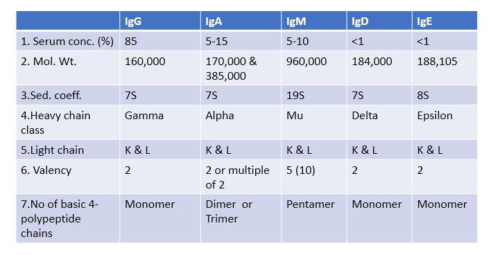
Properties of immunoglobulins


Overall Functions of Antibodies
- Based on antigen recognition and binding:-
1.mAb as B-cell receptor
2.sAb as antigen neutralizing agent
- Based on effector response:-
1. Complement activation
2. Opsonization
3. ADCC
- Based on Ig class:-
1. Neonatal immunity
2. Mucosal immunity
3. IgE mediated hypersensitivity reaction.
Antigenic determinant of Antibodies
- Antibodies are complex glycoproteins and they themselves can act as immunogens and induce antibody formation against them called anti-Ig antibodies.
- The whole Ig molecule is not immunogenic to the host system which produces antibodies to them, rather small sites or regions on the Ig molecule acts as immunogen.
- These sites or regions are called antigenic determinants.
- Based on their location on Ig, they are classified into three types:-isotypes, allotypes, and idiotypes.
- Isotypes:-formed by a unique sequence of amino acids located in the constant region of the H and L chain.
- Hence different isotypes differ from each other in their constant regions.
- Classes and subclasses of Igs are isotypes of one another.
- Isotypes are present in all the members of the same species and they are the same in all.
- Isotypes are different in different species. e.g. IgG of the mouse is different from the IgG of rabbit.
- The simplest way of producing isotype antibodies is to inject antibodies from one species to another.
Allotypes
- The antigenic determinant is present in the constant region of H and L chains and is encoded by polymorphic alleles, which are called allotypes.
- Since some members of a species carry alleles not all, they are present in some members of species.
- Allotypes differ in sequences of one to four amino acids from one another.
Idiotypes
- Are located in the hypervariable region of the VH and VL domains and one member of a species acts antigenic determinant to another member of the same species.
Polyclonal and Monoclonal antibodies
- An antigen usually has many epitopes and each epitope entering the body may stimulate a specific B-cell whose membrane receptor recognizes and binds to that epitope.
- So, many B-cells, each with unique specificity to one particular epitope are stimulated.
- Each stimulated B-cell produces antibodies specific to that one particular epitope.
- Therefore, the Serum of such an immunized subject contains a mixture of antibodies –specific to various epitopes present on an antigen.
- Such a mixture of antibodies is called polyclonal antibodies because it is derived from many B-cell clones contains Abs of different specificities.
- However only one B-cell is specifically stimulated by one particular epitope and is then allowed to proliferate and produce antibodies, then these Abs have two inherent characters: -They all are derived from one single B-cell and its clone. They all have the same antigenic specificity and such antibodies are called monoclonal antibodies.
- So monoclonal antibodies can be defined as the antibodies with identical antigenic specificity derived from a single B-cell clone.
- Monoclonal antibodies are produced by hybridoma technology.
Further Reading
- Essential immunology-Third Edition -Ivan M. Roitt
- Kuby Immunology –Sixth Edition-Thomas J. Kindt, Richard A. Goldsby, Barbara A. Osborne
- Basic Immunology –Second Edition -Abdul K. Abbas, Andrew H. Lichtman
- Immunology-Seventh Edition-Donald M. Weir, John Stewart
- Advances in Immunology- Volume-29 -F. J. Dixon, Henry G. Kunkel
- Fundamental Immunology-William E. Paul