
Trichophyton mentagrophytes: Introduction, Morphology, Pathogenecity, Laboratory Diagnosis and Keynotes
Introduction of Trichophyton mentagrophytes
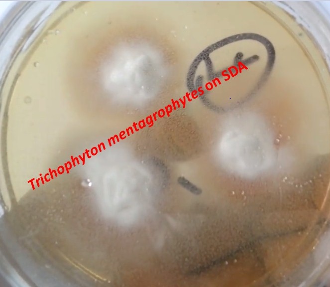
Trichophyton mentagrophytes colony characteristics on SDA are white to tan, cottony or powdery, pigment variables as shown above picture. Superficial fungal infections are a major global public health problem that affects 20–25% of the population worldwide. Among these diseases, dermatophytosis, or tinea, is one of the most frequent fungal infections. This infection is caused by dermatophyte species that belong to the Trichophyton, Microsporum, or Epidermophyton genera. T. mentagrophytes are filamentous fungi, digest keratin, and do not invade living tissues. They are incapable of penetrating subcutaneous tissue.
Morphology of Trichophyton mentagrophytes
This fungus is characterized morphologically based on the development of macro and microconidia with smooth walls. The colony is white to tan, cottony or powdery, pigment variables. The macroconidia originate laterally in the hyphae or in short pedicles of thin or thick walls and are club-shaped or fusiform, with a size that varies from 4-8 to 8-50 μm. The microconidia are abundant, spherical, pyriform, or irregularly shaped, with sizes varying from 2-3 to 2-4 μm. The most consistent feature of T. mentagrophytes is the production of globose micro-aleuriospores arranged in groups (like a bunch of grapes).
Pathogenecity of Trichophyton mentagrophytes
Dermatophytes are a group of fungi that are closely related to each other and have the enzyme keratinase; thus, they can cause infections in the skin, hair, and nails in both humans and animals. Among the dermatophytes, T. mentagrophytes stand out as the second most common causative agent of dermatophytosis after Trichophyton rubrum.
Mode of infection:- Acquired by direct contact with soil, animals, or humans infected with fungal spores.
Predisposing factors – Moist humid skin and tight-fitting underclothing.
Skin: grow in a centrifugal pattern in the stratum corneum annular or ring-shaped pruritic scaly skin lesions with central clearing and raised edges.
Nails: invade nails through lateral or superficial nail plates and then spread throughout the nails.
Hair shaft: Invade the hair shaft or maybe found surrounding it. Hairs become brittle and areas of alopecia may appear. Males more commonly infected as progesterone is inhibitory to dermatophyte growth.
Incubation period:- 1 to 2 weeks.
Anthropophilic dermatophytes:- commonest, cause relatively mild and chronic lesions but respond poorly to treatment.
Geophilic and zoophilic species:- less adapted to humans produce a more acute inflammatory response and severe infections; but they tend to resolve more quickly.
Clinical Types
Tinea capitis: Infection of the scalp (various types)
Kerion:- Painful inflammatory reaction producing boggy lesions on scalp Favus:- Cup like crust (scutula) forms around the infected hair follicle with minimal hair shaft involvement
Ectothrix:- Arthrospore formation occurs on the surface of the hair shaft
Endothrix:- Arthrospore formation occurs within the hair completely filling the hair shaft.
Tinea corporis: Infection of the non-hairy skin of the body e.g. trunk and limbs
Tinea pedis: Infect first the webs between the toes, then spread to the sole in a “moccasin” pattern. It is also called Athlete’s foot.
Tinea cruris ( Jock itch): Infection of the groin area
Tinea barbae: Infection of the beard and mustache area of the face
Tinea facie: Infection of the non-bearded area of the face
Tinea imbricate: Concentric lesions of the skin
Tinea unguium( nail plate infection): Infection of nail beds
Tinea manuum: Infection of the palmar aspect of hands
Laboratory Diagnosis of Trichophyton mentagrophytes
Specimens: It depends on the site of infection. e.g. Skin scraping in case of a skin infection, hair plucks for hair infection, and nail clipping from the active margin of the lesions of the nail
Transportation of specimen:-In moisture-free paper but when scrapings are to be sent through the post, they should be folded in thick black paper.
KOH Mount: branching septate hyaline mycelia, which frequently show arthrospore production. Hair- arthroconidia on the surface of the shaft (ectothrix) or within the shaft (endothrix). Septate hyaline hyphae and arthrospores of dermatophytes
Culture: Sabouraud dextrose agar (SDA) containing cycloheximide and incubated at 25°C, 30°C, and 37°C for 4 weeks. Potato dextrose agar – better sporulation (useful for the production of pigment). Colonies appear in 10 days to 3 weeks, depending on the organism strain. Dermatophytes test Medium (DTM)is used for presumptive identification of dermatophytes from fungal or bacterial contaminants found prevalent in cutaneous lesions. Incubation at 25°C.
Physiological characteristics
Urea hydrolyzation test: Positive
and hair perforation test: Positive
LPCB Preparation: LPCB stain stands for lactophenol cotton blue and it is a combination of fixative, staining, and clearing agent. LPCB uses both as a mounting fluid and a stain. This is used for staining and microscopic identification of fungi. Its contents functions are as follows- Lactic acid: It helps in preserving the morphology of the fungal elements. Phenol: It acts as a disinfectant. Cotton blue: It stains the fungal elements as well as intestinal parasitic (cyst, ova, and oocyst) and non-parasitic structures (vegetable cells, mucus, muscle fibers, and other artifacts). Glycerol: It is a hygroscopic agent that prevents drying. Tease the colony for LPCB mount which helps to demonstrate the hyphae and spore ( conidia). Conidia are of two types, microconidia -small unicellular while macroconidia are multicellular and septate. Special hyphae may have spiral hyphae, racquet hyphae, and favic chandeliers.
Molecular Identification: Internal transcribed spacer (ITS) region of the nuclear ribosomal DNA (rDNA) sequencing may be used to identify clinical species. The sequencing of a fragment from the region ITS1-5.8S-ITS2 of T. mentagrophytes complex was performed in the study of ‘ Molecular identification of isolates of the Trichophyton mentagrophytes complex’ in Mexico.
Skin test:-It detects hypersensitivity to trichophytin.
Treatment of dermatophytosis
Oral terbinafine or itraconazole is the drug of choice for the treatment of dermatophytosis.
Duration:- depends on the affected site (1 – 2 weeks for skin lesions, 6 weeks for hair infection, 3 months for onychomycosis)
They can be given as pulse therapy.
Alternative:- Oral griseofulvin and ketoconazole
Topical lotion:- Whitfield ointment or tolnaftate
Key Notes on Dermatohytes
- The taxonomy of T. mentagrophytes is complex due to the changes it has undergone in recent years. Until 2017, T. mentagrophytes-series included seven species: T. tonsurans, T. mentagrophytes, T. interdigitale, T, equinum, T. quinckeanum, T. schoenleinii, and T. simii characterized through ecological data, morphological characteristics, mating type studies, and molecular analysis.
- However, nowadays, only five species are considered—T. mentagrophytes, T. interdigitale, T. erinacei, T quinckeanum, and T. benhamie—as well as nine different genotypes of T. mentagrophytes / T. interdigitale associated with the geographical origin and the source of infection
- Conventionally, T. mentagrophytes is identified based on their macro and microscopic features, and sometimes, for their physiological characteristics i.e. hair perforation and urease activity.
- Woods Lamps Examination:- Positive for various Microsporum species and Trichophyton schoenleinii. Fluorescence is due to the presence of pteridine pigment in the cell wall.
- Key features of Trichophyton mentagrophytes are hyaline, septate, and branched hyphae as well as abundant spherical or semi-spherical microconidia that resemble clusters of grapes, spherical chlamyconidia, spiral hyphae, macroconidia, and nodular bodies.
- Trichophyton mentagrophytes and Microsporum canis are hair perforation test positive in which fungi pierce hair-producing wedge-shaped perforations.
Related Videos
#Ringworm and its causative agent dermatophyte under the microscope as shown below-
#Dermatophytes in KOH Preparation-
#Dermatophyte causing disease and its lab diagnosis |Trichophyton | Microsporum |Epidermophyton:
Dermatophytes are fungi that require keratin for growth. These fungi can cause superficial infections of the hair, skin, and nails consisting of three genus- Trichophyton, Microsporum, and Epidermophyton -spread by direct contact. Laboratory diagnosis on the following features -Site of infection, Colonial morphology, Presence of spores on LPCB tease mount- Microconidia and Macroconidia-
#Trichophyton mentagrophyte Isolated:
features- Helical pattern on LPCB Mount seen
Urease test-Positive
Hair perforation test-Positive
#Trichophyton rubrum- growth on dermatophyte test medium (DTM)
and its colonial morphology and LPCB tease mount under microscopy as shown in the video.
References
- https://www.hindawi.com/journals/ecam/2014/957860/
- https://www.ncbi.nlm.nih.gov/pmc/articles/PMC6945559/
- Textbook of Medical Mycology, Jagdish Chander.
- Essentials of Medical Microbiology, Apurba Sankar Sastry.
- https://www.slideshare.net/AnkurVashishtha4/dermatophytes
- https://www.researchgate.net/publication/327393673
- https://mycology.adelaide.edu.au/descriptions/dermatophytes/trichophyton/
- https://cmr.asm.org/content/cmr/8/2/240.full.pdf
- https://watermark.silverchair.com/46-8-811.pdf