
Tapeworm: Introduction, Morphology, Life Cycle, Clinical Features, Pathogenecity, Lab Diagnosis And Treatment
Cestode or tapeworm
Tapeworm is segmented, dorsoventrally compressed, and tape-like, therefore called tapeworm. It varies from a few millimeters to several meters in length. Adult cestode or tapeworm lives attached to the mucosa in the small intestine and absorbs food from the host intestine.
Scientific Classification of tapeworm
- Kingdom: Animalia
- Phylum: Platyhelminthes
- Class: Cestoda
- Order: Cyclophyllidea
- Family: Taeniidae
- Genus: Taenia
- Species: T. saginata
- T. saginata
Taenia saginata and Taenia solium
Taenia saginata : the beef tapeworm, the unarmed tapeworm.
Taenia solium : the pork tapeworm : the armed tapeworm of man.
T. saginata has a worldwide distribution in countries where cattle are raised and beef is eaten.
T. solium is not as widely distributed as T. saginata. It occurs mainly in Southern Africa, China, India, Central America, etc.
Habitat
Adult worms of both T. saginata and T. solium live in the small intestine (upper jejunum) of man.
Morphology of Taenia saginata
Adult worm
The adult worms consist of the scolex (head), neck, and strobila which is made of a large number of proglottids (segments).
- White and semitransparent
- Measures up to 10 meters
- Life span-10 years
- Morphology of proglottids (segments)
- 1000 to 2000 in number
- 2 cm long 0.5 cm wide
- Genital pore
- Marginal
- Posterior of segment
- Alternate right and left irregularity
- They are spherical, brown in color (bile stained), and measure 30-40 µm in diameter.
- They are surrounded by embryophore which is brown, thick-walled, and radially striated.
- Inside the embryophore, the hexacanth embryo (oncosphere) presents three pairs of hooklets.
- It does not float in a saturated solution of common salt (brine solution).
- It is viable for 8 weeks.
Morphology of Taenia solium
Adult worm
- Life span: 25 years
- 2-3 meters in length
- Adult worm contains three parts and they are-
Scolex
Neck: 0.5 mm
Proglottids
The genital pore is marginal.
Middle of the lateral margin of segments (alternating right and left)
Egg
30-40 µm
Spherical and brown in color
Thin outer transparent shell
Inner embryophore- brown radially striated
It contains an oncosphere with 3 pairs of hooklets.
It does not float in saturated saline solution.
Eggs are viable for up to 8 weeks.
Gravid segment
Size:12 mm ×6 mm
The longitudinal stem of the uterus with 5 to 10 lateral branches
Gravid segments are discharged passively in chains of 5 to 6 at a time
Cysticercus cellulosae (bladder worm)
Ovoid, opalescent ( 5 mm× 10 mm)
White vesicle
Covered by capsule and contain thick fluid 9 protein and salt)
The scolex of the larvae invaginated in the bladder
It is viable for many months.
It develops in men and pigs.
It becomes an adult worm in 5 to 12 weeks.
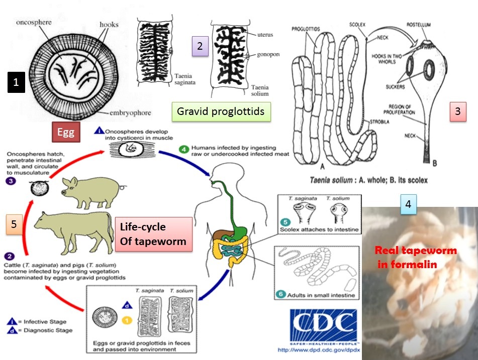
Life cycle of Taenia species
The T. saginata passes its life cycle in two hosts. A definitive host is a man who harbors the adult worm. The intermediate host is cattle. Eggs or gravid segments are passed out with the feces on the ground. These are ingested by cows or buffaloes.
Clinical features of Taenia saginata
Adult worms in the small intestine usually produce no symptoms. But at times, they may cause abdominal discomfort, indigestion, persistent diarrhea or diarrhea alternating with constipation and loss of appetite, intestinal obstruction, and even acute appendicitis.

Clinical features of Taenia solium
Vague abdominal discomfort
Indigestion
Alternating diarrhea and constipation
Anemia
Cysticercus cellulose infection
Ingestion of eggs in water or vegetables
autoinfection
Solitary or multiple
Commonly subcutaneous and muscles
Any other organs may be involved
Eyes
- Blurring vision
- Uveitis
- Iritis
- Loss of vision
Brain
Epilepsy
Behavioral disorder
Paresis
Hydrocephalus
Less often organs are-
- Heat
- Liver
- Lung
- Abdominal cavity
- Spinal cord
- Brain
Cysticercus cellulosae is surrounded by fibrous capsules except for eye and brain ventricles
Evokes cellular response
Pathogenicity
Cysticercosis is a disease caused by the larval stage of T. solium, an important public health problem in tropical countries. Occasionally obstructive appendicitis or cholangitis occurs in Taenia infections due to aberrant migration of segments, which have been found also in the uterine cavity. In rare cases, proglottids may obstruct the respiratory tract, enter the middle ear through the eustachian tube or localize in adenoid tissue of the nasopharynx. They usually develop in the subcutaneous tissues and muscles forming visible nodules. It may develop in the brain.
Laboratory diagnosis
Specimens: Stool, a biopsy of subcutaneous nodules, blood, serum
Direct method
Demonstration of egg and gravid segments.
By microscopy method; saline wet mount: Saline wet mount is made by mixing a small quantity (about 2 mg of feces) in a drop of saline placed on a clean glass slide. Remove any gross fibers or particles and cover with a coverslip. The smear is then examined under a microscope.
Concentration methods: Formalin-ether sedimentation; for species diagnosis- head and gravid segments are required. It can’t be differentiated morphologically.
Anal swab: Anal swabs are superior to the methods using for detecting eggs from feces.
The diagnosis of cysticercosis can be carried out using biopsy of the subcutaneous nodule, it may reveal Cysticercus cellulosa.
Indirect method
X-ray of skull and soft tissue to reveal calcified cysticerci. MRI for non-calcified cyst
Blood cell count: eosinophilia
Serological test for detection of specific antibodies by using serum, CSF, IHA
(indirect hemagglutination) , IFA (Indirect fluorescent antibody) and ELISA.
Treatment
Praziquantel and niclosamide can be used for the treatment of human tapeworm infection. Other drugs are-
- Quinacrine
- Bithionol
- Mebendazole
- for cysticercosis
- surgical excision
- Praziquantel
- Metrifonate
Prophylaxis of T. saginata infection
All beef to be eaten by man should be inspected for cysticerci. Through cooking of beef ensures complete protection.
Proper sanitary disposal of feces.
Cattle should not be allowed to feed or graze on the ground polluted by human feces or sewage. This will control cattle infection.
Prophylaxis of T. solium infection
Personal hygiene.
General sanitary measures.
Avoid food and water contamination with T. solium eggs.
A strict veterinary inspection of pork in all slaughterhouses with condemnation of infected pigs. Thorough cooking of pork ensures complete protection.
Further Readings
- Medical Parasitology by Abhay R. Satoskar, Gary L. Simon, Peter J. Hotez and Moriya Tsuji
- Atlas of Medical Helminthology and protozoology -4th edn -P.L. Chiodini, A.H. Moody, D.W. Manser
- Merkel and Voge’s medical parasitology
9th edition. - Parasitology: 12th edition
By K. D. Chatterjee - District laboratory practice in Tropical countries –Part-I.
By Monica Chesbrough. - Isenberg clinical microbiology procedures Handbook
2nd edition. Vol. 2 - Atlas of Human Parasitology, Lawrence R Ash, Thomas C. Orihel, 3 rd ed, Publisher ASCP Press, Chicago.
- Topley & Wilson’s Principle of parasitology. Editors: M.T. Parker & amp; L.H. Collier, 8 th ed 1990, Publisher Edward Arnold publication, London.
- Molecular Medical Parasitology. Editors: J. Joseph Marr, Timothy W. Nilsen, and Richard W. Komuniecki, Publisher Academic Press, an imprint of Elsevier Science.