
Hydrogen Sulfide Production by Various Organisms and Its Detection Methods
Hydrogen Sulfide Production by Various Organisms
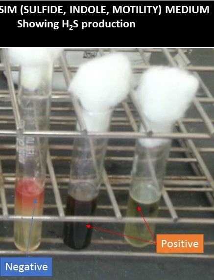
Hydrogen sulfide production by various organisms i.e. Proteus mirabilis and Salmonella Typhi from left to right as shown above image.
H2S of bacteria can be detected by the following tests-
- Triple sugar iron (TSI) agar test
- SIM (Sulfide, Indole, Motility ) medium test
Introduction of TSI test
This single TSI test is useful for the following features-
- To know the carbohydrate utilization ability of the organisms
- Gas formation by the organisms
- Hydrogen sulfide production by the bacteria
TSI stands for triple sugar iron and it is agar medium recommended for use in the differentiation of Enterobacteriaceae by their ability to ferment glucose, lactose, and sucrose, and their ability to produce hydrogen sulfide.
Principle of TSI test
Triple sugar iron agar contains casein and meat peptones, phenol red as the pH indicator, 0.1% glucose, 1% lactose, and 1% sucrose for fermentation. Ferric or ferrous ions and sodium thiosulphate are present to detect hydrogen sulfide production. Bacteria that are non lactose fermenting initially produce a yellow slant due to the production of acid from the glucose. The small amount of glucose is rapidly depleted. Oxidation metabolism continues in the slant after the low concentration of glucose has been depleted, producing an alkaline pH from the aerobic breakdown of peptone; the slant turns red. There is no oxygen penetration into the butt and no oxidative metabolism; the butt remains acid and yellow. Thus, a non lactose fermenting bacterium yields an alkaline (K) slant over an acid (A) butt ( K/A; red slant; yellow butt). Lactose/sucrose bacteria continue to produce a large amount of acid in the slant and in the butt so the reaction in both remains acid (A/A; yellow slant; yellow butt). If the slant and butt remain neutral, the organism is not capable of fermenting glucose or other sugars (K/K; red slant; red butt) Bubble, fracturing, or displacement of the medium indicates gas production by the organism due to sugar fermentation. Blackening of the medium denotes hydrogen sulfide production by the action of the bacteria with sodium thiosulfate and which is detected by the reduction of the ferric ions to produce a black precipitate.
Requirements for TSI test
- Test organism
- Triple sugar iron agar
- Bunsen burner and inoculating needle
- BOD incubator
- Test tubes rack
- QC strains for quality control
Test Procedure
- Warm medium to room temperature and examines for cracks and don not use if cracks appear.
- Touch the center of a well-isolated colony by using a sterile inoculating needle.
- Stab to within 3-5 mm from the bottom of the tube.
- Place cap loosely on tube
- Incubate aerobically at 35-37°C for 18-24 hours.
Observation and Interpretation
Examine the reaction in the slant and the butt also observe for gas and hydrogen sulfide production.
- Yellow: Acid reaction
- Red: Alkaline reaction
- Blackening of the medium: H2S production
- Bubbles, cracks, or displacement of the medium: Gas production
Interpretation of carbohydrate
- A/A: Glucose and lactose or sucrose fermented
- K/A: Only glucose fermented or non-lactose fermenter
- K/K: No carbohydrate fermented or non-glucose fermenter
Result
Tube No 1
- K/K
- No H2S production
- No gas formation
Tube No 2
- R/A
- H2S positive
- No gas formation
Tube No 3
- A/A
- Gas formation positive
- No H2S formation
Limitations of TSI test
- Do not read the TSI test before 18 hours, since false readings of acid in the slant may result.
- H2S production may on be inhibited on the TSI test for organisms that utilize sucrose and suppress the enzyme mechanism that results in the production of H2S.
- Sulfide indole motility (SIM) agar is more sensitive in the detection of H2Sthan either TSI or KIA.
Key Notes
- Kligler’s Iron Agar(KIA) test only differs from the Triple Sugar Iron agar (TSI) test due to lacking sucrose in its composition.
- Do not attempt to interpret sugar fermentation reactions after 24 hours. Refrigerate tubes if reading will be delayed.
- If desired, extend incubation only to detect H2S production. Campylobacter may take 3 days for the production of H2S.
Introduction of SIM test
SIM (Sulfide, Indole, Motility ) medium is useful for the differentiation of gram-negative enteric bacilli. SIM test helps to isolate the organisms on the basis of sulfide production, indole formation, and motility.
Principle of SIM test
The medium having the constituents ferrous ammonium sulfate and sodium thiosulfate, which together serve as indicators for the production of hydrogen sulfide (H2S). Hydrogen sulfide production detects when ferrous sulfide, a black precipitate, is produced as a result of ferrous ammonium sulfate reacting with hydrogen sulfide gas. Casein peptone of this medium is rich in tryptophan. Organisms having the enzyme tryptophanase degrade tryptophan to indole. Indole detection is achieved after the addition of Kovac’s reagent following incubation of the inoculated medium. Indole combines with p-dimethylaminobenzaldehyde and produces a red band at the top of the medium. A negative indole test produces no color change after the addition of Kovac’s reagent i.e. Yellow color of Kovac’s reagent. A lower concentration of agar added to the medium provides a semi-solid structure allowing for the detection of bacterial motility. Motile organisms diffuse from the stab line and produce turbidity or cloudiness throughout the medium. The growth of non-motile bacteria is restricted along the stab line and leaves the surrounding medium clear. Another constituent, animal tissue of this medium which provides amino acids and nutrients necessary for bacterial growth.
Composition of the medium
Ingredients for 100 ml of distilled water-
Pancreatic Digest of Casein: 2.0gm
Peptic Digest of Animal Tissue: 0.61gm
Ferrous Ammonium Sulfate: 0.02gm
Sodium Thiosulfate: 0.02gm
Agar: 0.35gm
Final pH 7.3 +/- 0.2 at 25°C.
Requirements for SIM test
- Test organisms
- SIM medium
- Inoculating wire and
- Bunsen burner
- Incubator
Quality control strains
- Escherichia coli ATCC 25922
- Salmonella enterica ATCC 14028
Procedure of SIM test
- Take pure colonies from an 18-24-hour old culture on solid medium.
- Inoculate the SIM Medium by stabbing the center of the medium to a depth of half inch.
- Incubate the inoculated medium aerobically at 37°C for 18-24 hours.
- Observe for hydrogen sulfide production and motility of test organism.
- Only apply Kovac’s reagent (three drops ) after reading the result of H2S and motility reaction to the surface of the medium.
- Observe for the development of a pink to red color.
Result interpretation of SIM test
- Positive H2S test: blackening of the medium
- A negative H2S test: absence of blackening
- Positive motility test: a diffuse zone of growth flaring from the line of inoculation
- Negative motility test: restricted growth along the stab line
- Indole positive test: a pink to red color ring is formed at the top of the medium after the addition of Kovac’s reagent
- Indole negative test: A yellow color denotes a negative indole test after the addition of Kovac’s reagent
- Escherichia coli ATCC 25922: Growth; Motility: positive, H2S: negative, and Indole: positive (It turns pink after addition of Kovac’s reagent)
- Salmonella enterica ATCC 14028: Growth; Motility: positive, H2S: positive, and Indole: negative
Limitations of SIM test
- Caps should be loose during incubation otherwise erroneous results may occur
- The inoculum should take from a solid medium because a liquid or broth suspension will delay the initiation of growth and may cause erroneous results.
- When inoculating semi-solid media, it is important that the inoculating needle be removed along the exact same line used to inoculate the medium. A fanning motion may result in growth along the stab line that may result in false-positive interpretation.
- Take motility and hydrogen sulfide (H2S) reaction results prior to addition of Kovac’s reagent.
- Weakly motile organisms or organisms that possess damaged flagella (due to heating, shaking, or other trauma) often result in false-negative motility tests and therefore, motility results should be confirmed by performing a hanging drop motility test.
- Some microorganisms like Yersinia enterocolitica demonstrates motility best at 25°C.
- Obligate aerobes, such as Pseudomonas aeruginosa , will produce a spreading film on the surface of the medium and will not extend from the line of inoculation where oxygen is depleted.
Further Readings
- Cowan & Steel’s Manual for identification of Medical Bacteria. Editors: G.I. Barron & R.K. Felthani, 3rd ed 1993, Publisher Cambridge University Press.
- Bailey & Scott’s Diagnostic Microbiology. Editors: Bettey A. Forbes, Daniel F. Sahm & Alice S. Weissfeld, 12th ed 2007, Publisher Elsevier.
- Clinical Microbiology Procedure Hand book, Chief in editor H.D. Isenberg, Albert Einstein College of Medicine, New York, Publisher ASM (American Society for Microbiology), Washington DC.
- Colour Atlas and Text book of Diagnostic Microbiology. Editors: Koneman E.W., Allen D.D., Dowell V.R. Jr and Sommers H.M.
- Jawetz, Melnick and Adelberg’s Medical Microbiology. Editors: Geo. F. Brook, Janet S. Butel & Stephen A. Morse, 21st ed 1998, Publisher Appleton & Lance, Co Stamford Connecticut.
- Mackie and Mc Cartney Practical Medical Microbiology. Editors: J.G. Colle, A.G. Fraser, B.P. Marmion, A. Simmous, 4th ed, Publisher Churchill Living Stone, New York, Melborne, Sans Franscisco 1996.
- Text book of Diagnostic Microbiology. Editors: Connie R. Mahon, Donald G. Lehman & George Manuselis, 3rd edition2007, Publisher Elsevier.