
Yeast cells in Gram stain stained smear: Lab diagnosis of Superficial Mycoses
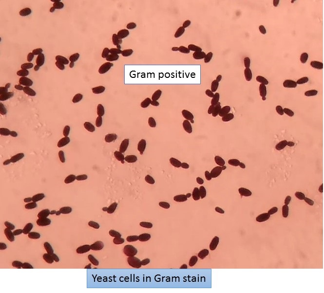
Yeast cells in Gram stain
Yeast cells of Candida albicans from SDA agar in gram stain showing oval shape Gram positive as shown above picture.
Yeast cells found in following fungi-
Candida species
Cryptococcus species
Dimorphic fungi in parasitic phase –
Blastomyces dermatitidis
Histoplasma capsulatum
Coccidioides immitis
Paracoccidioides brasiliensis
Sporothrix schenckii
Penicillum marneffei
Laboratory Diagnosis Of Superficial Mycose
Superficial mycoses is the infection of superficial layer i.e. keratinized layers of the skin and its appendages and mucosa of the oral cavity and vagina in case of mucosal candidiasis. Etiology – surface mycoses is caused by Malassezia furfur, Exophiala werneckii, Trchosporon beigelli and Piedraia hortae and cutaneous mycoses is caused by dermatophytes and in case of mucosal candidiasis by Candida spp.
Sample collection for superficial mycoses
Samples may be obtained by scraping the skin and nails with a scalpel blade or microscope slide holding at 90°. Skin samples are scraped from the outer edges of the surface lesions.Skin and nail must be cleaned with 70 % isopropanol before sampling. Infected hair should be removed by plucking with forceps and never by cutting because cutting fails to remove the area most likely to harbor the fungus i.e. the base of the hair shaft around the follicle. The collection of hair sample is facilitated by wood’s lamp. In case of onychomycosis, the patient should stop anti-fungal agents one week prior to the collection of specimen. Nail specimens should be clipped from the free edge. The fungus in the distal portion of nail is often non-viable & if culture does fail samples can be taken from the base of nail. In case of superficial candidiasis the sample is collected by repeatedly rubbing the swab firmly over the white plaques.
Skin stripping
This is an alternative method for the collection of skin specimen. A waterproof transparent adhesive tape is applied firmly to the affected area and peel it off. The tape now bearing a thin layer of skin is then applied to the sterile glass slide with KOH and observe for fungal elements i.e. fungal hyphae or yeast cells.
Hair brush sampling technique
Hair sample may be collected from scalp by brushing with a sterile plastic hair brush or scalp massage pad which is then inoculated into culture medium by pressing the brush or pad spines into the SDA.
Storage and transport of sample for superficial mycoses
These samples should be allowed to dry out because the moisture causes to overgrow the contaminants. Black paper containing the specimen may be folded to form a packet. In such conditions ringworm fungi remain viable for weeks or even months(up to 12 months). Specimen should not be refrigerated as viability of some species(Epidermophyton spp.) is affected. The survival of yeast cells on dry swab is inversely related to time and storage temperature. In case of delay can be stored at 4⁰C .
Direct Examinations
Direct examination of clinical material provides a rapid report to the physician to initiate the treatment. In some cases specific morphological characteristics provide a clue to the genus of organisms. It may provide evidence of infection despite negative cultures in case of anti-fungal therapy.
Microscopic examination
KOH preparation
Principle- The fungal cell wall glucans being alkali resistant, is not dissolved in KOH and other skin layer, keratin, mucus and other tissue components are dissolved.
10 or 20 or 30 % KOH is used. Skin, nail, hair or other samples obtained are mixed with a drop of KOH on a slide and covered with a cover slip. Heat may be applied to potentiate the clearing of tissue. If the specimen is transparent it may be examined without treatment with KOH. Malassezia furfur shows oval or bottle shaped yeast cells along with short, curved hyphae. Hortae werneckishows brownish, branched, septate hyphae and budding cells. Trichosporon beigelli shows hyphae and rectangular arthrospores within(endothrix) and around(exothrix) hair. Piedraia hortae shows dark color septate hyphae around hair and asci containing 2-8 aseptate ascospores. Dermatophytes in skin scraping, nail clippings or nail scraping and hair stubs can appear as refractile, hyaline, septate, branched or unbranched hyphae and arthrospores. Several modifications of the basic 10% KOH preparation have been made for more rapid detection. This includes incorporating Parker superchrome blue in the KOH solution for selective staining of the fungal elements like hypahe and yeast cells.
Other modifications of the basic methods includes
- Addition of 36% of DMSO to 20% KOH to clearing of the specimen without heating.
- Addition of 5-10% glycerine to the KOH preparation to delay crystallization of the KOH, degrading of the fungus and dehydration.
- In KOH preparation it is very difficult to differentiate ‘mosaic fungus’ (cholesterol crystals deposited around the periphery of the epidermal cells). It can be recognized by lack of internal organelles.
- Parker’s ink can also be used for better visualization of Malassezia furfur and (Scopuloropsis spp. and Candida spp.) causing onychomycosis.
KOH with calcofluor white
Calcofluor white is a whitening agent used in the textile and paper industry. A drop of calcofluor white (a fluorescent dye) can be added to the KOH preparation before adding a cover slip. Calcofluor white binds to the polysaccharide present in the chitin or to cellulose. Fungal element fluoresce apple green or blue white. So, any element with a polysaccharide skeleton will fluoresce. Although background elements may also fluoresce, the fungal components are brighter.
Culture
Common solid media are as SDA, or DTM with antimicrobial agent and cycloheximide. Small fragments of keratinous material are planted or scattered on SDA containing chloramphenicol and cycloheximide and incubated at room temperature. Growth is slow and colonies may appear in 1-3 weeks. Specialized media may be necessary to stimulate sporulation such as cornmeal agar, potato flake agar, lactrimel rice grains or PDA so that sporulation will occur. Culture from suspected superficial mycoses is incubated at 30ºC. DTM incorporates gentamicin and chloramphenicol to inhibit the bacteria and a phenol red indicator that changes from yellow to red when the medium becomes alkaline as the result of growth of dermatophytes.
Identification of culture
Colony characteristics(macroscopy)- Gross colony features observed on SDA include color of the surface, color of reverse, texture of the surface(powdery, granular, velvety or fluffy) type of folding(radial, cerebriform) and the rate of growth.
Microscopic examination of culture
Microscopic characteristics that should be observed are the following:
Septate or aseptate hyphae, hyaline or dematiaceous hyphae, fruiting structures, the types, size, shape and arrangement of conidia. Microscopic examinations can be studied in teased mounts, slide cultures or cellophane tape preparation.
Tease mount
With the help of two teasing needles, a portion of the mycelium is removed.
A LPCB preparation is prepared on a slide. LPCB does not work well with the dematiaceous fungi because they retain their dark color. The major disadvantage of this procedure is the disruption of conidia during the teasing process.The preparations may be mounted in Poirrer’s blue or lactophenol aniline blue or carbol fuschin .
Cellophane tape preparation
Cellophane tape preparations involve gently touching a piece of clear tape, sticky side down to the surface of the colony and then removing it. The tape is placed onto a drop of LPCB on a slide and examined. With this procedure the conidial arrangement is retained.
Slide culture
Slide cultures are useful for demonstrating the natural morphology of the fungal structures and for encouraging conidiation in some poorly fruiting fungi. Slide culture is particularly useful when the slide culture from a known isolate is stored in a collection for future comparison against isolates awaiting identification.
Slide culture Technique
Culture character and microscopy of dermatophytes
Reference
- Medical Mycology. Editors: Emmons and Binford, 2nd ed 1970, Publisher Lea and Febiger, Philadelphia.
- Rippon’s JW: Medical Microbiology. The pathogenic fungi and the pathogenic Actinomycetes. 3rd ed 1988 Publisher WB saunder co, Philadelphia.
- Clinical Microbiology Procedure Hand book Vol. I & II, Chief in editor H.D. Isenberg, Albert Einstein College of Medicine, New York, Publisher ASM (American Society for Microbiology), Washington DC.
- A Text Book of Medical Mycology. Editor: Jagdish Chandar. Publication Mehata, India.
- Practical Laboratory Mycology. Editors: Koneman E.W. and G.D. Roberts, 3rd ed 1985, Publisher Williams and Wilkins, Baltimore.
- Topley & Wilsons Medical Mycology. Editors: M.T. Parker & L.H. Collier, 8th ed 1990, Publisher Edward Arnold publication, London.
- Text book of Diagnostic Microbiology. Editors: Connie R. Mahon, Donald G. Lehman & George Manuselis, 3rd edition2007, Publisher Elsevier.
- Mackie and Mc Cartney Practical Medical Microbiology. Editors: J.G. Colle, A.G. Fraser, B.P. Marmion, A. Simmous, 4th ed, Publisher Churchill Living Stone, New York, Melborne, Sans Franscisco 1996.
- Bailey & Scott’s Diagnostic Microbiology. Editors: Bettey A. Forbes, Daniel F. Sahm & Alice S. Weissfeld, 12th ed 2007, Publisher Elsevier.