
Trichosporon LPCB Preparation: Introduction, Pathogenecity, Lab Diagnosis and Treatment
Trichosporon LPCB preparation
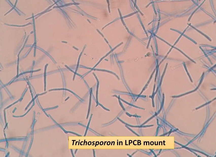
Trichosporon LPCB preparation as shown above image has hyaline, septate hyphae that fragment into oval or rectangular arthroconidia.
Introduction Of Trichosporon
The fungus, Trichosporon, genus is characterized by the development of hyaline, septate hyphae that fragment into oval or rectangular arthroconidia. Few blastoconidia are also visible. The colonies are usually raised and have a waxy appearance, that develops furrows and irregular folds as shown above picture. Most common species, especially from invasive infections and comes under risk group -2 organism.
Pathogenicity
6 species of medical importance are listed below-
- Trichosporon asahii
- Trichosporon asteroides
- Trichosporon cutaneum
- Trichosporon inkin
- Trichosporon mucoides and
- Trichosporon ovoides.
- They are a minor component of normal skin flora and are widely distributed in nature. They are regularly associated with the soft nodules of white piedra, and have been involved in a variety of opportunistic infections in immunosuppressed patients. Disseminated infections are most frequently caused by T. asahii and have been associated with the following conditions like leukemia,
- organ transplantation
- multiple myeloma
- aplastic anemia
- lymphoma
- solid tumors and
- AIDS.
Disseminated infections are often fulminated and widespread, with lesions occurring in the liver, spleen, lung, and gastrointestinal tract. Infections in non-immunosuppressed patients include endophthalmitis after surgical extraction of cataracts, endocarditis usually following insertions of prosthetic cardiac valves, peritonitis in patients on continuous ambulatory peritoneal dialysis (CAPD), and intravenous drug abuse.
Laboratory Diagnosis
Trichopsoron species can be diagnosed in the laboratory using the following techniques-
- KOH mount of specimens
- Cultivation of specimens in fugal media like SDA, PDA, etc
- Observation of fungal growth on LPCB preparation under the microscope
- Assimilation tests
Treatment
Following anti-fungal drugs are useful-
- Fluconazole
- Itraconazole
- Posaconazole
- Voriconazole
- Amphotericin B
- Flucytosine
- Caspofungin
- Anidulafungin
Further Readings
- Medical Mycology. Editors: Emmons and Binford, 2nd ed 1970, Publisher Lea and Febiger, Philadelphia.
- Description of Medical Fungi, Editors: David Ellis, Stephen Davis, Helen Alexiou, Rosemarry Handake, Robyn Bartley, 2nd edition
- Rippon’s JW: Medical Microbiology. The pathogenic fungi and the Pathogenic Actinomycetes. 3rd ed 1988 Publisher WB Saunder co, Philadelphia.
- Clinical Microbiology Procedure Handbook Vol. I & II, Chief in editor H.D. Isenberg, Albert Einstein College of Medicine, New York, Publisher ASM (American Society for Microbiology), Washington DC.
- A Textbook of Medical Mycology. Editor: Jagdish Chander. Publication Mehata, India.
- Practical Laboratory Mycology. Editors: Koneman E.W. and G.D. Roberts, 3rd ed 1985, Publisher Williams and Wilkins, Baltimore.