
LE Cell in Blood Smear: Definition, Introduction, Pathophysiology and Lab Diagnosis
Definition of LE Cell
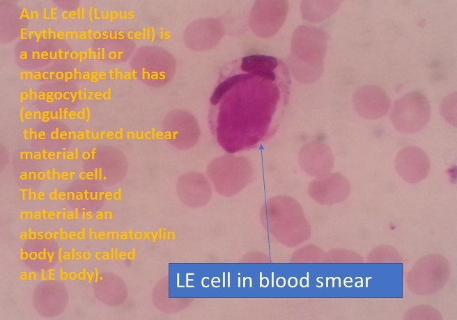
An LE cell is a neutrophil or macrophage that has phagocytized the denatured nuclear material of another cell. The denatured material is an absorbed hematoxylin body and it is also called an LE body. LE stands for Lupus Erythematosus.
Introduction of LE Cell
LE cell in blood smear as shown above image. It is useful to diagnose Systemic lupus erythematosus (SLE) in which 5 mL of venous blood of the patient is taken and is traumatized by a glass rod or glass beads. Positive LE Cells present in Systemic lupus erythematosus (95% of the cases), Drug-induced lupus erythematosus (80 to 95 % of the cases) and Other autoimmune diseases show LE cell in less than 20 % of the cases.
Pathophysiology of LE Cell
SLE is a typical autoimmune disease and is a systemic rheumatic disease. Etiology is unknown but the primary defect is in the immune system. 5 to 7 new cases are diagnosed per year per 100000 people. The range of the disease is mildly limited to a photosensitive facial rash and transient diffuse arthritis. Life-threatening involvement of kidneys, cardiovascular system, respiratory system, central nervous system, etc. In SLE there are various self-antigens like nuclear histone and proteins, immunoglobulins G ( IgG), red blood cells( RBCs), DNA and RNA, and platelets. Developed autoantibodies form against all the above antigens may give rise to a complex of antigen and antibody (Ag+Ab) which will lead to damage by Type 3 and Type 2 hypersensitivity reaction. Antinuclear antibodies cause damage to the nucleus which degenerates and gives rise to a homogeneous body called a hematoxylin body.
Laboratory Diagnosis of LE Cell
Requirements
- Centrifuge tubes, glass beads, rubber bungs
- Clean and grease-free microscopic glass slides
- Leishman’s stain
- Centrifuge
- Microscope
- Vortex mixer or blood mixing rotor
- Specimen: 5 ml EDTA blood
Test Procedure
- Take about 5 ml of EDTA blood in a test tube (15 x 125 mm)
- Add five glass beads and fit the stopper of the tube.
- Rotate on a blood mixing rotor at 50 RPM for 30 minutes.
- Place the tube at 37°C for 10 minutes.
- Transfer the blood to a Wintrobe tube.
- Centrifuge Wintrobe tube at 3000 RPM for 30 minutes.
- Remove hemolysed plasma and then remove carefully the buffy coat.
- Make smears of the buffy coat on clean grease-free and dry glass slides.
- Fix the air-dried smears in methanol.
- Stain with Leishman stain.
- Screen under high power(40X) and observe specific areas under the oil immersion objective.
Observation of stained smears
- Le cell appears as a neutrophil containing a large spherical body in its cytoplasm as shown above picture.
- The LE body does not show nuclear structure and stains as a pale purple homogenous mass.
Result
LE cell: Present
Report
LE cells: Positive
Keynotes on LE Cell
- Lupus erythematosus (LE) cell testing has been replaced by antinuclear antibody (ANA) testing.
- The tart cells retain their nuclear structure and should not be confused with LE cells.
Bibliography
- https://www.annualreviews.org/doi/abs/10.1146/annurev.me.11.020160.001311?journalCode=med
- A Test Book of Medical Laboratory Technology-Praful B. Godaka and Darshan P. Godkar, 2nd Edition
- https://www.labpedia.net/…
- https://emedicine.medscape.com/article/2087069
- https://www.sciencedirect.com/topics/immunology-and-microbiology/le-cell
- https://www.ncbi.nlm.nih.gov/pmc/articles/PMC6211931/