
Exserohilum: Case Report, Classification, Morphology, Pathogenecity and Lab Diagnosis
Exserohilum Case Report
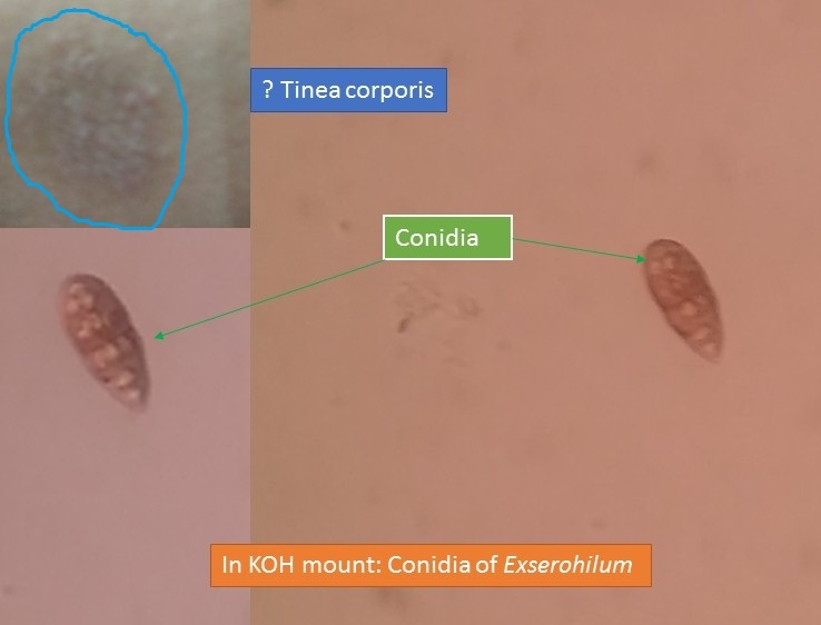
- Exserohilum conidia in potassium hydroxide mount of Tinea corporis suspected site as shown above picture.
- Exserohilum is a dematiaceous fungus and also an agent of phaeohyphomycosis.
- After the use of 1% clotrimazole ointment, a patient is a free form of fungal infection.
- Again performing potassium hydroxide preparation of that infected site, no fungal elements were seen.
Scientific Classification
- Kingdom: Fungi
- Division: Ascomycota
- Class: Dothideomycetes
- Order: Pleosporales
- Family: Pleosporaceae
- Genus: Exserohilum
- species: Exserohilum rostratum
Note: Clinically important species are E. rostratum and
E. longirostratum.
Habitat and Distribution
Exserohilum has a cosmopolitan distribution and is found naturally in warm, tropical, and subtropical locations on grasses, rotten wood, and in the soil.
Morphology
Exserohilum in potato dextrose agar (PDA) and produce woolly colonies that are pale initially but quickly become dark gray, to bluish-black to brownish-black with a black reverse. Hyphae are septate and brown. Conidiophores may be up to 200 µm long, are septate, non-branched, geniculate, and become paler near the apex. Conidia are various shades of brown and may range from less than 80 to greater than 100 µm in length, maybe straight to distinctly curved, and contain 1 to over 12 septa. The feature all species share is a strongly protruding hilum from the basal cell. Conidia may be slow to form in culture.
Pathogenicity of Exserohilum
Exserohilium is one of the causative agents of phaeohyphomycosis. It causes
- cutaneous infection in immunocompromised patients
- Subcutaneous phaeohyphomycoses
- keratitis as well as a fatal disseminated infection in a patient with aplastic anemia and
- corneal ulcer
Lab Diagnosis
Specimens
Direct examination of specimens
- Macroscopy
- Microscopy (KOH preparation, staining techniques, direct immunofluorescence, and histology)
Culture and isolation
- Identification of culture growth
Colony character
Microscopy ( Tease mount, cellophane tape preparation, and slide culture)
Nucleic acid probes - An indirect method based on host immune response
Skin testing
Serological tests
Miscellaneous tests for the identification of yeasts and molds.
Reference
- Medical Mycology. Editors: Emmons and Binford, 2nd ed 1970, Publisher Lea and Febiger, Philadelphia.
- https://drfungus.org/knowledge-base/exserohilum-species/
- Rippon’s JW: Medical Microbiology. The pathogenic fungi and the Pathogenic Actinomycetes. 3rd ed 1988 Publisher WB Saunder co, Philadelphia.
- A Textbook of Medical Mycology. Editor: Jagdish Chander. Publication Mehata, India.
- Practical Laboratory Mycology. Editors: Koneman E.W. and G.D. Roberts, 3rd ed 1985, Publisher Williams and Wilkins, Baltimore.