
Dermatophyte Growth on SDA: Introduction, Culture, Microscopic Examination and Lab Diagnosis
Introduction of Dermatophyte
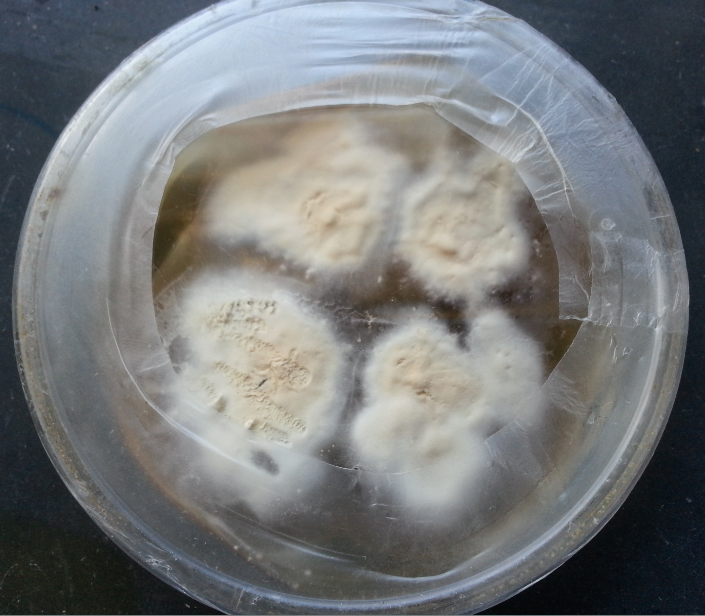
Dermatophyte is a group fungus of three genera Trichophyton, Microsporum, and Epidermatophyton. This one is Trichophyton mentagrophyte powdery pigmented colonies on Sabouraud dextrose agar (SDA). It causes ringworm.
Culture of Dermatophyte
Common solid media are as SDA, or DTM with antimicrobial agent and cycloheximide. Small fragments of keratinous material are planted or scattered on SDA containing chloramphenicol and cycloheximide and incubated at room temperature. Growth is slow and colonies may appear in 1-3 weeks. Specialized media may be necessary to stimulate sporulation such as cornmeal agar, potato flake agar, lacrimal rice grains, or PDA so that sporulation will occur. Culture from suspected superficial mycoses is incubated at 30ºC. DTM incorporates gentamicin and chloramphenicol to inhibit the bacteria and a phenol red indicator that changes from yellow to red when the medium becomes alkaline as the result of the growth of dermatophytes.
Identification of Culture
Colony characteristics(macroscopy)- Gross colony features observed on SDA include the color of the surface, the color of reverse, texture of the surface(powdery, granular, velvety, or fluffy) type of folding (radial, cerebriform), and the rate of growth.
Microscopic examination of culture for dermatophyte
Microscopic characteristics that should be observed are the following:
Septate or aseptate hyphae, hyaline or dematiaceous hyphae, fruiting structures, the types, size, shape, and arrangement of conidia. Microscopic examinations can be studied in teased mounts, slide cultures, or cellophane tape preparation.
Tease Mount
With the help of two teasing needles, a portion of the mycelium is removed. An LPCB preparation is prepared on a slide. LPCB does not work well with the dematiaceous fungi because they retain their dark color. The major disadvantage of this procedure is the disruption of conidia during the teasing process. The preparations may be mounted in Poirier’s blue or lactophenol aniline blue or carbol fuchsin.
Cellophane Tape Preparation of Dermatophyte
Cellophane tape preparations involve gently touching a piece of clear tape, sticky side down to the surface of the colony, and then removing it. The tape is placed onto a drop of LCB on a slide and examined. With this procedure, the conidial arrangement is retained.
Slide Culture of Dermatophyte
Slide cultures are useful for demonstrating the natural morphology of the fungal structures and for encouraging conidiation in some poorly fruiting fungi. Slide culture is particularly useful when the slide culture from a known isolate is stored in a collection for future comparison against isolates awaiting identification.
Slide Culture Technique
Culture character and microscopy of dermatophytes
Physiological And Biochemical Tests For Fungal Infections Laboratory Diagnosis
- Hair perforation test
Trichophyton rubrum, which may be morphologically similar to T. mentagrophytes, usually causes only surface erosion of hair shafts in this test, whereas T. mentagrophytes usually form perpendicular pegs in the hair shafts. This test can also be used to distinguish penetration capable Microsporum canis from M. equinum which does not penetrate the hair. - Urease test
5 days urease test help to differentiate T. mentagrophytes from T. rubrum. Tubes of Christensen urea agar are very lightly inoculated with the dermatophytes and held for 5 days at room temperature. Most isolates of T. mentagrophytes demonstrate urease production resulting in a color change. Most T. rubrum isolates are negative or require more than 5 days to give a positive reaction. - Growth on rice grains
This test is useful to differentiate poorly sporulating isolates of M. canis from M. audouinii. Sterile nonfortified rice is inoculated lightly with a portion of a colony. After 10 days of incubation at room temperature, the medium is observed for growth. M. canis and other dermatophytes grow well and usually form many conidia whereas M. audouinii does not grow but rather turns the rice grains brown.
Further Readings
- Medical Mycology. Editors: Emmons and Binford, 2nd ed 1970, Publisher Lea and Febiger, Philadelphia.
- Rippon’s JW: Medical Microbiology. The pathogenic fungi and the Pathogenic Actinomycetes. 3rd ed 1988 Publisher WB Saunder co, Philadelphia.
- Clinical Microbiology Procedure Handbook Vol. I & II, Chief in editor H.D. Isenberg, Albert Einstein College of Medicine, New York, Publisher ASM (American Society for Microbiology), Washington DC.
- A Textbook of Medical Mycology. Editor: Jagdish Chander. Publication Mehata, India.
- Practical Laboratory Mycology. Editors: Koneman E.W. and G.D. Roberts, 3rd ed 1985, Publisher Williams and Wilkins, Baltimore.
- Topley & Wilsons Medical Mycology. Editors: M.T. Parker & L.H. Collier, 8th ed 1990, Publisher Edward Arnold publication, London.
- Textbook of Diagnostic Microbiology. Editors: Connie R. Mahon, Donald G. Lehman & George Manuselis, 3rd edition2007, Publisher Elsevier.
- Mackie and Mc Cartney Practical Medical Microbiology. Editors: J.G. Colle, A.G. Fraser, B.P. Marmion, A. Simmous, 4th ed, Publisher Churchill Living Stone, New York, Melborne, Sans Franscisco 1996.
- Bailey & Scott’s Diagnostic Microbiology. Editors: Bettey A. Forbes, Daniel F. Sahm & Alice S. Weissfeld, 12th ed 2007, Publisher Elsevier.