
Aspergillus fumigatus Colony Characteristics: Introduction, Pathogenecity, Laboratory Diagnosis, Treatment and Prevention
Aspergillus fumigatus Colony Characteristics
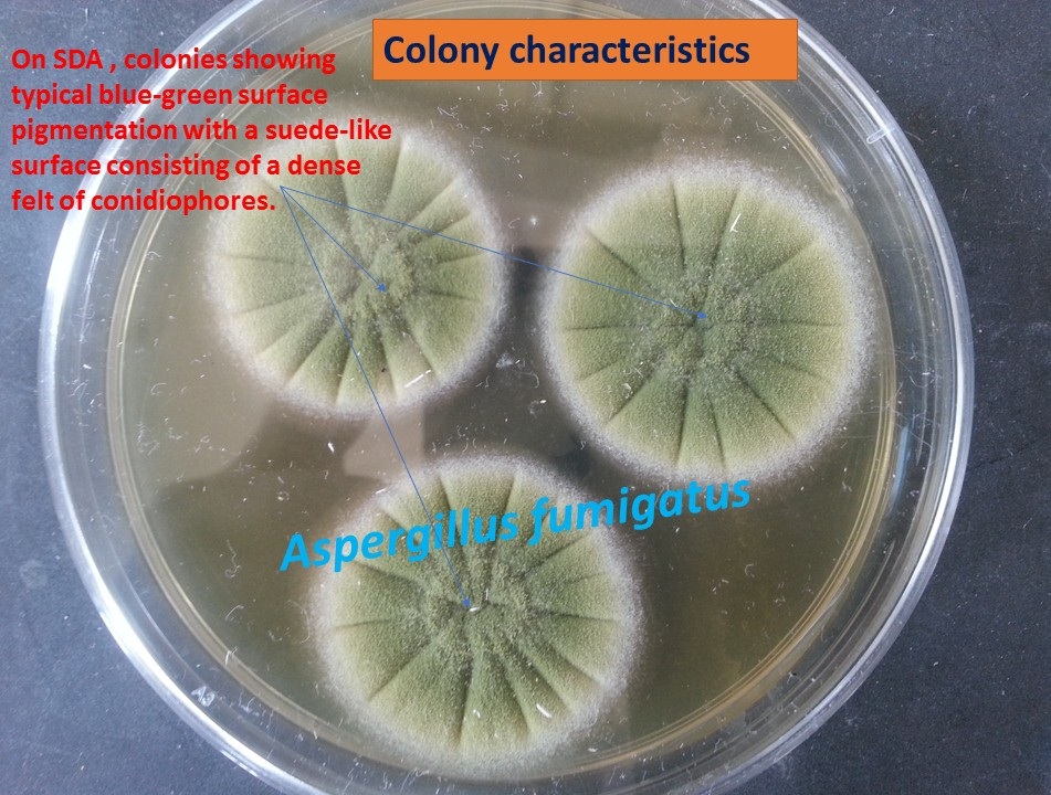
Aspergillus fumigatus colony characteristics as shown above picture. It is characterized by green echinulate conidia, 2.5 to 3 μm in diameter, produced in chains basipetal from greenish phialides, 6 to 8 by 2 to 3 μm in size. A few isolates of these organisms are pigment less and produce white conidia. The chains of conidia are borne directly on broadly clavate vesicles (20 to 30 μm in diameter) in the absence of metulae . No sexual stage is known for this species. A. fumigatus is a fast grower; the colony size can reach 4 ± 1 cm within a week when grown on Czapek-Dox agar at 25°C and this is also a compatible feature for SDA as shown above figure ( after 4 days of incubation). It is a thermophilic species, with growth occurring at temperatures as high as 55°C and survival maintained at temperatures up to 70°C.
Key Features of Aspergillus fumigatus
Uniseriate and columnar conidial head with the phialides limited to the upper two-thirds of the vesicle and curving to be roughly parallel to each other.
Introduction of Aspergillus fumigatus
A. fumigatus is one of the most ubiquitous of the airborne saprophytic fungi and opportunistic pathogen. We constantly inhale numerous conidia of this fungus. The conidia are normally eliminated in the immunocompetent host but complications in immunosuppressed or immunocompromised patients causing fungal infections like allergic bronchopulmonary aspergillosis, chronic pulmonary aspergillosis, aspergilloma, and even invasive aspergillosis (IA).
Classification of Aspergillus fumigatus
(Micheli -1729)
- Kingdom: Fungi
- Division: Ascomycota
- Class: Eurotiomycetes
- Order: Eurotiales
- Family: Trichocomaceae
- Genus: Aspergillus
- Species: Aspergillus fumigatus
Pathogenicity
A. fumigatus can cause the following infections-
- allergic bronchopulmonary aspergillosis,
- chronic pulmonary aspergillosis,
- aspergilloma and
- invasive aspergillosis.
Allergic Bronchopulmonary Aspergillosis
This condition is an allergic reaction to the Aspergillus spores. This reaction can lead to damage to your airways and lungs. It is more prone to people who have asthma and cystic fibrosis.
Chronic pulmonary aspergillosis
It develops progressively and can occur in people with chronic lung conditions that cause air spaces called cavities to form in the lung. e.g. tuberculosis and emphysema. It can be manifested by following ways-small spots of Aspergillus infection in the lungs, called nodules tangled balls of fungus within a lung cavity, called aspergillomas (these can sometimes cause complications such as bleeding in the lungs) more widespread infection of multiple lung cavities, which may or may not contain aspergillomas When left untreated, the widespread infection can lead to thickening and scarring of lung tissue, which can lead to loss of lung function.
Aspergilloma
An aspergilloma is a clump of mold which exists in a body cavity such as a paranasal sinus or an organ such as the lung. Aspergilloma mainly affects people with underlying cavitary lung diseases such as tuberculosis, sarcoidosis, bronchiectasis, cystic fibrosis, and systemic immunodeficiency.
Invasive Aspergillosis
Invasive aspergillosis is the most severe form of aspergillosis. It can be fatal if not treated. It occurs when an aspergillosis infection begins in the lungs and spreads to other parts of your body like skin, brain, or kidneys. IA occurs only in people who have a severely weakened immune system.
Symptoms of Invasive Aspergillosis
The symptoms of invasive aspergillosis may contain-
- fever
- cough, which can include coughing up blood
- shortness of breath
- chest pain, which can be worse when you take deep breaths
Note: When the infection spreads outside of the lungs, symptoms may depend on which part of the body is affected, but may include symptoms like as follows-
- headache
- nosebleed
- swollen eyes
- lesions on the skin
- joint pain
- difficulties with speech
- confusion
- seizures
Risk Factors
- Blood cancers
- Asthma
- Cystic fibrosis
- Neutropenia
- Long-term corticosteroid therapy
- Organ transplant
- Patient on immunosuppressive drugs
- AIDS
Laboratory Diagnosis of Aspergillus fumigatus
Specimen: It depends on the site of infection even though common specimens may be sputum, bronchial wash, tracheal aspirates collected from patients with pulmonary disease. Tissue biopsies from patients with disseminated disease. Ear discharge is from chronic suppurative otitis media (CSOM) patient.
Direct Microscopy: Using potassium hydroxide ( KOH) mount or calcofluor white, sputum specimens, bronchoalveolar lavage (BAL), bronchial biopsies show non-pigmented septate hyphae with repeated dichotomous branching as shown below.
#Fungal elements (septate hyphae with dichotomous branching) in-ear discharge-
Cultural isolation: Colonies are typically blue-green with a suede-like surface consisting of a dense felt of conidiophores as shown above picture.
LPCB Preparation of Fungal Growth: Uniseriate and columnar conidial head with the phialides limited to the upper two-thirds of the vesicle and curving to be roughly parallel to each other.
#Aspergillus fumigatus Colony on SDA, LPCB tease mount under microscopy as shown in video-
Antigen Detection: There are several antigen tests for the detection of Aspergillus from blood, urine, and CFS. In a person with invasive aspergillosis, there may be high levels of galactomannan antigen in serum. Galactomannan is rapidly eliminated from the blood, therefore serial screening twice weekly is recommended for optimal diagnosis.
Serology: Immunodiffusion tests for the detection of antibodies to Aspergillus species in the diagnosis of allergic, aspergilloma, and invasive aspergillosis. Patients with Allergic Bronchopulmonary Asthma have high levels of IgE specific for Aspergillus antigens and some IgG precipitins.
Molecular Identification: Sequence analysis of ITS is sufficient to identify to species complex level only. For definitive identification analysis, β-tubulin, calmodulin, and actin genes are required (Samson et al. 2007; Balajee et al. 2005).
Other supportive Methods of Diagnosis: A chest X-ray to look for signs of infection, such as aspergillomas.
Treatment of Aspergillus fumigatus Infections
Treatment depends on the site of involvement. e.g. Allergic bronchopulmonary aspergillosis can be treated with oral corticosteroids, similar chronic pulmonary aspergillosis that consists of nodules or single aspergilloma may not require treatment if you don’t have any symptoms. The nodules should be monitored regularly to make sure that they don’t progress. Whereas antifungal medications are used to treat more serious cases of chronic pulmonary aspergillosis, as well as invasive aspergillosis using antifungal agents voriconazole, itraconazole, and amphotericin B. Nowadays resistance of A. fumigatus to azole antifungal medications is also reported and they are voriconazole and itraconazole. In this case, amphotericin B is the choice of drug. A surgical removal is also an option if aspergillomas are causing complications such as bleeding in the lungs.
Keynotes
- Aspergillus was first viewed under a microscope in 1729 by the Italian priest and biologist Pier Antonio Micheli.
- Environmental surveys indicate that everyone will inhale at least several hundred A. fumigatus conidia per day.
- An A. fumigatus infection can be difficult to diagnose because the symptoms often resemble other lung conditions such as tuberculosis.
Prevention and Control
A. fumigatus are present throughout the environment. For this reason, it can be difficult to prevent exposure. However, in this scenario, there are some steps that can make infection less likely.
- Avoid activities that are more likely to bring you into contact with A. fumigatus like involvement in gardening, yard work, or visiting construction sites otherwise wear long pants and sleeves and wear gloves if you’ll be handling soil or manure and use an N95 respirator when you are in very dusty areas.
- Bone-marrow transplant units should have filtered air-conditioning systems, monitored airborne contamination of patients, reduced visits to patients, and application of measures that isolate patients and minimize the risks of exposure to Aspergillus conidia and molds.
- Take prophylactic antifungal medication. e.g. If you’ve recently undergone a procedure like an organ transplant, your doctor may prescribe antifungal medications to prevent infection.
- If you’re in an at-risk group, periodic testing for Aspergillus may help to detect an infection in its early stages.
- If you’re in a group that’s at risk for developing aspergillosis, share with your doctor. They can tell you ways that you can prevent becoming infected.
- Preventive measures like prophylactic low dosage of amphotericin B or Itraconazole for persons at risk.
References
- https://cmr.asm.org/content/33/1/e00140-18
- https://mycology.adelaide.edu.au/descriptions/hyphomycetes/aspergillus/
- https://www.ncbi.nlm.nih.gov/pmc/articles/PMC88920/
- https://www.healthline.com/health/aspergillus-fumigatus
- https://en.wikipedia.org/wiki/Aspergillus
- https://www.immunology.org/public-information/bitesized-immunology/pathogens-and-disease/aspergillus-fumigatus
- https://ec.asm.org/content/6/11/1953
- https://www.sciencedirect.com/topics/medicine-and-dentistry/aspergillus-fumigatus