
Blood Parasites: Introduction, Pathogenecity, Lab Diagnosis and Treatment
Blood Parasites
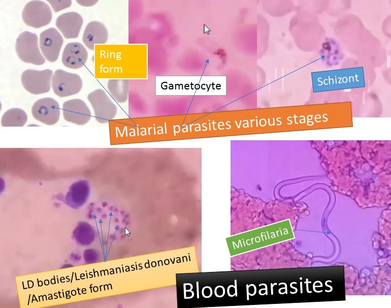
Blood parasites like a malarial parasite, Leishmania donovani, and microfilaria and their structures as shown above picture. Details of these organisms are as follows.
Introduction of Malarial Parasites
Malarial parasites are a mosquito-borne infectious agent that causes a disease called Malaria. These agents are Plasmodium falciparum, P. vivax, P. ovale, and P. malariae and commonly called malarial parasites. These four species of malaria parasites infect us. All are transmitted by female Anopheles mosquitoes. According to a report, 3.3 billion people are at risk of infection, malaria remains one of the world’s most significant health problems. P. falciparum is the most common species identified (nearly 75%) followed by P. vivax (nearly 20%) responsible for causing malaria. A new emerging species, Plasmodium knowlesi is a zoonotic species that causes malaria in macaques and these are mostly of limited public health importance.
Classification of malarial parasites
Kingdom: Animalia
Sub kingdom: Protozoa
Phylum: Protozoa
Class: Sporozoa
Genus: Plasmodium
Species: vivax, falciparum, ovalae and milariae
Geographical distribution – malaria is present worldwide in tropical and subtropical areas.
Mode of Transmission
Naturally acquired infections are via the bite of infected female Anopheles mosquitoes. Malaria is also transmitted via blood transfusion, sharing of contaminated needles among IV drug abusers, and congenital transmission also has been documented.
Vector: Transmitted by over 60 species of female Anopheles mosquito. The male mosquito feeds exclusively on fruits and juices. The female needs at least 2 blood meals, before the first batch of eggs, can be laid.
Life cycle of malarial parasites
Definitive host: Female Anopheles mosquito
Intermediate host: Man
The malaria parasite multiplies by the division or splitting of a process known as schizogony.
A female Anopheles mosquito transmits a motile infective form (called the sporozoite) to humans (the secondary host), thus acting as a transmission vector. A sporozoite travels through the blood vessels to liver cells (hepatocytes), where it reproduces asexually (tissue schizogony), producing thousands of merozoites. These infect new red blood cells and initiate a series of asexual multiplication cycles (blood schizogony) that produce 8 to 24 new infective merozoites, at which point the cells burst and the infective cycle begins anew. Whereas other merozoites develop into immature gametocytes, which are the precursors of male and female gametes. When a fertilized mosquito bites an infected person, gametocytes are taken up with the blood and mature in the mosquito gut. The male and female gametocytes fuse and form an ookinete—a fertilized, motile zygote. Ookinetes develop into new sporozoites that migrate to the insect’s salivary glands, ready to infect a new host. The sporozoites are injected into the skin, in the saliva, when the mosquito takes a subsequent blood meal.
Pathogenicity of this blood parasite
Malaria infection develops via two phases: one that involves the liver (exoerythrocytic phase), and one that involves erythrocytes (erythrocytic phase). When an infected mosquito pierces a person’s skin to take a blood meal, sporozoites in the mosquito’s saliva enter the bloodstream and migrate to the liver where they infect hepatocytes, multiplying asexually and asymptomatically.
Acute Symptoms
Classical features include cyclic symptoms
Cold stage: chills and shaking
Hot stage: fever, headache, vomiting, seizures in children
Sweating stage: weakness
Feel well for a period of time, then the cycle repeats itself
Other Physical symptoms:
Fever: Fever can be very high from the first day. Temperatures of 40°C and higher are often observed. Fever is usually continuous or irregular. Classic periodicity may be established after some days. Hepatomegaly: The liver may be slightly tender. Splenomegaly: Splenomegaly takes many days, especially in the first attack in nonimmune children. In children from an endemic area, huge splenomegaly sometimes occurs. Anemia: Prolonged malaria can cause anemia, and malarial anemia causes significant mortality.Jaundice: With heavy parasitemia and large-scale destruction of erythrocytes, mild jaundice may occur. This jaundice subsides with the treatment of malaria. Dehydration: High fever, poor oral intake, and vomiting all contribute to dehydration.
Each disease has a distinct course
“Tertian Malaria” (P. falciparum, P. ovale, and P. vivax)fever occurs every third day. “Quartan Malaria” (P. malariae)fever occurs every fourth day. P. ovale and P. vivax can cause chronic malaria, reappearing after months or years due to latent parasites in the liver.
Laboratory Diagnosis of malarial parasites
- Demonstration of Parasite by Microscopy
Diagnosis of malaria can be made by demonstration of malarial parasite in the blood. Thick smears: It is recommended that 200 oil immersion fields should be examined before a thick film is declared negative.Thin smears: for detecting the parasites and determining the species.
- Quantitative Buffy Coat, Smear
RBC containing malaria parasites is less dense and concentrate just below the buffy coat of leucocytes at the top of the erythrocytic column. Pre-coating of the tube with acridine orange induces a fluorescence on the parasites, which can be visualized under oil immersion. The nucleus of the parasite appears as fluorescing greenish-yellow against a red background.
- Micro concentration Technique
A blood sample is collected in a microhematocrit tube and centrifuged at high speed. The sediment is mixed with normal serum and a smear is prepared. Though it increases the positivity rate, it changes the morphology of the parasite.
- Culture of Malaria Parasites
Several culture lines have been established from the blood of infected Aotus monkeys or directly from human patients. Schizogony proceeds normally in culture. Gametocytes are formed infrequently. Plasmodium retains its infectivity in culture.
- Serodiagnosis:
Serodiagnosis is not helpful in clinical diagnosis because it will not differentiate between an active and past infection. It is used mainly for a seroepidemiological survey and to identify the infected donors in transfusion malaria. The tests used are indirect hemagglutination (IHA), indirect fluorescent antibody test (IFA), and enzyme-linked immunosorbent assay (ELISA).
- Rapid Antigen Detection Tests:
Based on the detection of antigens using the immunochromatographic method. Have been developed in different test formats like the dipstick, card, and cassette-bearing monoclonal antibody, directed against the parasite antigens.
- Fluorescence Microscopy
Fluorescent dyes like acridine orange or benzothiocarboxy purine are used, which stain the parasites entering the RBCs but not WBCs. This is a method of differential staining. Acridine orange stains DNA as fluorescent green and cytoplasmic RNA as red.
- Molecular Diagnosis
The DNA probe is a highly sensitive method for the diagnosis of malaria. It can detect less than 10 parasites/µL of blood. Polymerase chain reaction (PCR) is increasingly used now for species specification and for the detection of drug resistance in malaria.
- Other Tests
Measurement of hemoglobin and packed cell volume (PCV)Total WBC and platelet count in severe falciparum malaria.
Measurement of blood glucose to detect hypoglycemia in severe falciparum malaria and patients receiving quinine Coagulation tests like measurement of anti-thrombin III level, plasma fibrinogen, fibrin degradation products (FDPs), prothrombin time (PTT), if abnormal bleeding is suspected in falciparum malaria.
Urine for free hemoglobin, if blackwater fever is suspected.
Blood urea and serum creatinine to monitor renal failure.
Treatment of Malarial Parasites
Chloroquine (Aralen)
Quinine sulfate (Qualaquin)
Hydroxychloroquine (Plaquenil)
Mefloquine combination of atovaquone and proguanil (Malarone)
Control of Malaria
It can control using the following methods-
- Using medicine for disease control-Medicine prepares from silicon tree, bark-quinin,e and trade names- Chloroqunin, Mepaquinin
- by mosquito control
It is further of three types-
- Physical control
- Chemical control and
- Biological control ( Use of Gambasia fish)
Introduction of Leishmaniasis
Leishmaniasis is an infectious disease caused by the unicellular blood parasite, Leishmania. Members of the genus Leishmania pass their life cycle in two hosts- man and the insect vector female sandfly. On the basis of clinical disease production, it is of various types like Visceral leishmaniasis, Cutaneous leishmaniasis, and cutaneous and mucocutaneous leishmaniasis. So, here we discuss visceral leishmaniasis in detail and it is also called Kala-azar, which is the most severe form of Leishmaniasis.
Classification of Leishmaniasis
Classification of Leishmaniasis ( based on disease production )
- Visceral leishmaniasis
Leishmania donovani (complex) (VL): L. donovani, L. Infantum, and L. chagas - Cutaneous leishmaniasis of the Old world:
Leishmania tropica
Leishmania major
- Cutaneous and mucocutaneous leishmaniasis of New world
Leishmania mexicana (Complex)
Leishmania brazilliensis (complex)
Leishmania peruriana
Leishmania donovani
It causes visceral leishmaniasis or kala-azar and also causes post-kala-azar dermal leishmaniasis.
Geographical Distribution
India ( states- Assam, Bengal, Bihar, Orissa, UP)
China
Africa
South America
South Europe
Russia
Habitat: Amastgote form in the reticuloendothelial system (macrophages): e.g. spleen, bone marrow, liver, etc.
Transmission: Persons to person transmitted by the bite of female sand flies (Phlebotomus or Lutzomyia).
Morphology of Leishmania donovani
It has two forms promastigote and amastigote.
Promastigote
It is long slender, spindle-shaped about 15-20 µm length, 1-2µm width.
The nucleus is centrally placed.
Kinetoplast (blepharoplast and parabasal body ) lies transversely and near the anterior end.
Flagellum projects from the anterior end and may be of the size of the body or even longer.
Habitat: a digestive tract of insect vector (sandfly) or laboratory culture.
Amastigote
Amastigote form is also called LD bodies.
It has shape and size: round or oval body, 2-4 micrometer along the longitudinal axis.
Kinetoplast (parabasal body and the blepharoplast ) at a right angle to the nucleus.
Axoneme arises from blepharoplasty and extends up to the tip of the cell.
Vacuole alongside the axoneme is present.
Habitat: It is present in reticuloendothelial cells of the body.
Life cycle of Leishmania donovani
The life cycle of Leishmania completes in two hosts, human, and sandfly. Sandfly is a bloodsucker, usually feeding at night on sleeping prey. When the fly bites a leishmaniasis patient, the pathogen is ingested along with the prey’s blood. Inside the stomach of the sandfly, the amastigotes quickly transform into elongated and motile forms called the promastigotes. The promastigotes live extracellularly in the alimentary canal, reproducing asexually, then migrate to the proximal end of the gut. As the fly bites, the promastigotes are released from the proboscis and introduced locally at the bite site. Once in our body, promastigotes invade macrophages where they transform back into the smaller amastigote form. The amastigotes replicate in the macrophage cell, inside the phagolysosome but only when whose normal defensive response they are able to prevent. The daughter cells then migrate to fresh cells or through the bloodstream to find new hosts. In this way the infection is progressive, spreading to our body’s mononuclear phagocyte system, particularly the spleen and liver. The free amastigotes in peripheral tissues are then ingested by sandfly to start another cycle.
Pathogenesis of Visceral Leishmaniasis
Infections range from asymptomatic to progressive, fully developed visceral leishmaniasis/ kala-azar. The incubation period is 3-6 months. Progressive wasting, anemia, and protrusion of the abdomen from enlarged liver and spleen. Fatal after 2 – 3 years if not treated. In acute cases with chills, fevers up to 104°F, and vomiting; death may occur within 6 – 12 months. The immediate cause of death is usually an invasion of a secondary pathogen that the body is unable to combat.
Clinical Manifestation of Visceral Leishmaniasis
Pyrexia- Low-grade fever ( continuous intermittent in later stages)
Hepato-splenomegaly
Characteristic blackening of skin with dry, rough, harsh
Anemia
Leukopenia
Cachexia
Hypergammaglobulinemia
Epistaxis
Proteinuria,
Hematuria
Hair ( brittle, fallout)
Post-Kala-azar Dermal Leishmaniasis
Its beief form is PKDL. Normally it develops <2 years after completion of antimonial treatment for the original disease when the visceral infection disappears but the skin infections persist.
Clinical presentation of PKDL
Hypopigmented patches,
Erythematous patches
Yellowish pink nodules
Laboratory Diagnosis of Leishmaniasis
- Direct evidence:
Specimens: Bone marrow biopsy, spleen or liver biopsy (rare ), Blood (common)
Microscopy: Leishman’s/wright or Giemsa stain
Isolation: Culturing of promastigote form in NNN (Novy-Nicolle-McNeal medium)
This medium contains defibrinated rabbit blood (one part) and salt agar ( 2 parts ).
After inoculating the material in the water of condensation of the medium, it is incubated at 22°C to 24°C.
After completion of incubation, it demonstrates promastigote form of parasites.
Schneider’s insect tissue culture medium
Animal inoculation: e.g. Golden hamster
Ag detection: e.g. Immunoblotting and PCR
- Indirect evidence
Blood picture: leucopenia, anemia
Napier’s Aldehyde test
Chopra’s Antimony test
Complement fixation test with WKK antigen-non specific as the antigen is prepared from tubercle bacillus by Witebsky, Klingenstein, and Kuhn.
Immunofluorescence test
Indirect Hemagglutination Test
Qualitatively detects anti-leishmania circulating antibodies by leishmanial recombinant antigen K39 (rK39)- a product of a gene cloned from L. chagasi containing a 39- aminoacid repeat conserved among Leishmania species.
Rapid immunochromatographic tests
Leishmanin Skin Test
0.2 ml of killed culture intradermal injection- positive in cured cases of Kala-azar and negative result in active leishmaniasis.
Treatment of Leishmaniasis
Standard treatment
Pentavalent antimonial sodium stibogluconate intravenously
Pentamidine intramuscularly (used if fail to respond antimonial sodium stibogluconate )
Amphotericin B may be used in the pentamidine resist cases.
Control of Leishmaniasis
Vector control
Reservoir control
Treatment of active cases
Vaccination
Cutaneous Leishmaniasis
Its causative agent is Leishmania tropica.
Pathogenesis
Infected sandfly injects promastigote form
The core of the cells parasitized by amastigote form
Acanthosis cellular infiltration
Pressure necrosis and ulceration
Secondary infection
Granulation
Healing with a scar takes 2 to 12 months.
Cutaneous and mucocutaneous Leishmaniasis
Its causative agent is Leishmania brazilliensis.
Pathogenesis
Infected sandfly injects promastigote form.
Weeping ulcer (oriental sore)
Spreads to the mucosa of mouth, nose, larynx, pharynx
Parasitized cells inflammatory infiltration necrosis
later on reactive fibrosis
Introduction of Filariasis
Filariasis is an infectious disease caused by nematodes of the Filarioidea type e.g. Wuchereria bancrofti, Brugia malayi, Oncocerca volvulus, and Loa loa. These are spread by blood-feeding insects such as black flies and mosquitoes. The filarial worms reside in the subcutaneous tissues, lymphatic system, or body cavities of humans,
Classification of filarial worms based on location in the body
- lymphatic filariasis – causative agents: Wuchereria bancrofti, Brugia malayi, Brugia timorii,
- Subcutaneous filariasis: Causative agents are-Loa loa, Oncocerca volvulus, Mansonella streptocerca
- Serous cavity filariasis: Causative agents are-Mansonella perstans, Mansonella ozzardi (They are virtually non-pathogenic)
Microfilaria
The females of parasites are viviparously giving birth to larvae so-called microfilaria. This parasite includes four genera and species that are parasitic to humans. These filarial nematodes are as follows: 1. Wuchereria bancrofti 2. Brugia malayi 3. Oncocerca volvulus and 4. Loa loa but mainly two genera i.e. Wuchereria and Brugia are encountered in peripheral blood.
Wuchereria bancrofti
Geographical Distribution
- India
- Japan
- South china
- South America
- Central and West Africa
Habitat
Lymphatics and lymph nodes
Morphology
It has two stages adult worm and larva (microfilaria).
Adult worm
The adults are whitish, translucent, thread-like worms with smooth cuticles and tapering ends. The female is larger (70–100 × 0.25 mm) than the male (25–40 × 0.1 mm). The posterior end of the female worm is straight, while that of the male is curved vertically and contains 2 spicules of unequal length. Males and females remain coiled together usually in the abdominal and inguinal lymphatics and in the testicular tissues. The female worm is viviparous and directly liberates sheathed microfilariae into the lymph.
Lifespan: 10 to 15 years
Microfilaria
The microfilaria has a colorless, translucent body with a blunt head, and pointed tail It measures 250–300 µm in length and 6–10 µm in thickness
It is covered by a hyaline sheath, within which it can actively move forwards and backward Along the central axis of the microfilaria, a column of granules can be seen, which are called somatic cells or nuclei. The granules are absent at certain specific locations—a feature that helps in the identification of the species. Microfilariae do not multiply or undergo any further development in the human body. Their lifespan is believed to be about 2–3 months. It is estimated that a micro filarial density of at least 15 per drop of blood is necessary for infecting mosquitoes
Periodicity
The microfilariae circulate in the bloodstream. They show a nocturnal periodicity in peripheral circulation; being seen in large numbers in peripheral blood only at night (between 10 pm and 4 am). This correlates with the night-biting habit of the vector mosquito.
Life Cycle
Definitive host: Man
Intermediate host: Female mosquito, of different species, acts as vectors in different geographic areas. The vector in most other parts of Asia is Culex quinquefasciatus (C. fatigans).
Infective form: Actively motile third-stage filariform larva
Mode of transmission: Bite of a mosquito carrying filariform larva.
- Microfilaria in peripheral capillaries ( at night )
- Microfilaria ingested by mosquito reaching the stomach
- Microfilaria, shed sheath, penetrate the gut wall, enter thoracic muscles
- Short larvae ( first stage )
- Second stage larvae
- Third stage larva
- Mosquito bites man and depositing of infective larva on skin
- Larva skin penetration, lymphatics and becomes adult male and female
- Adult worms male and female remain coiled together ( abdomen, inguinal lymphatics, testicular tissues) and again cycles starts
Clinical features of Filariasis ( due to Wuchereria bancrofti )
Clinical features of filariasis differ according to the stages of the worm and the nature of its location in our body.
Due to larvae entry into the blood
- Malaise
- Headache
- Nausea
- vomiting
- Low-grade fever
- Pruritis
- Urticaria
- Fugitive swelling ( skin or limb)
Occult Filariasis
- Due to hypersensitivity to filarial antigen
- Microfilaria is not detectable in blood but seen at affected sites
- May present as tropical pulmonary eosinophilia
- Fever
- Loss of weight
- Anorexia
- Dry nocturnal cough
- Dyspnea
- Asthmatic wheezing
- blood eosinophil count 3000 to 5000 cu mm
- May also show
- Glomerulonephritis
- Arthritis
- Tenosynovitis
- Thrombophlebitis
- Dermatosis
Adult worm
- Lymphangitis (testicles, epididymis, spermatic cord, etc)
- Lymphadenitis (groin, axilla)
- Hydrocele
- Elephantiasis
- Chyluria
Laboratory Diagnosis of Filariasis
- Demonstration of Microfilaria: Microfilaria can be demonstrated in blood, chylous urine, exudate of lymph varix, and hydrocele fluid. Peripheral blood is the specimen of choice. The method has the advantage that the species of the infecting filaria can be identified from the morphology of the microfilaria seen.
- Unstained Film: Examination under the low-power microscope shows the actively motile microfilariae lashing the blood cells around. The timing of blood collection is critical and should be based on the periodicity of the microfilariae.
- Concentration Techniques: When the microfilaria density is low, concentration techniques are used: Knott’s concentration technique
Nucleopore filtration
DEC provocation test
- Biopsy: Adult filarial worms can be seen in sections of biopsied lymph nodes, but this is not employed in routine diagnosis
- Skin Test: Intradermal injection of filarial antigens induces an immediate hypersensitivity reaction. But the diagnostic value of the skin test is very limited due to the high rate of false-positive and negative reactions.
- Serodiagnosis: Complement fixation, indirect hemagglutination (IHA), indirect fluorescent antibody (IFA), immunodiffusion, and immune enzyme tests have been described. But the tests are not sufficiently sensitive or specific to be used either for individual diagnosis or surveys. Demonstration of Circulating Antigen Highly sensitive and specific test for detection of specific circulating filarial antigen (CFA)The Trop bio test is a semiquantitative ELISA for detection of CFA in serum or plasma specimen. Immunochromatographic filariasis card test (ICT) is a new and rapid filarial antigen test that detects soluble W. bancrofti antigens using a monoclonal antibody (ADIZ) in the serum of infected humans. Both assays have sensitivities of 93–100% and specificities approaching 100%.
- Molecular Diagnostic Technique: Polymerase chain reaction (PCR) can detect filarial DNA from a patient’s blood, only when circulating microfilaria are present in peripheral blood but not in chronic carrier state usually the test provides sensitivities that are up to 10 fold greater than parasitic detection by direct examination and is 100% specific.
- Other tests:
Indirect Evidence
Eosinophilia (5–15%) is a common finding in filariasis.
Elevated serum IgE levels can also be seen.
Treatment of Filariasis
Diethylcarbamazine (DEC): effective against adult and microfilariae. Ivermectin: destroy microfilariae but not adults.
Vector Control
Drainage and filling:
urban chemical and biological larvicides
Indoor residual insecticide
spraying
Outdoor residual insecticide spraying
Personal protection
Insecticide
impregnated materials: nets, curtains, clothing
House screening
House location
Repellents
Fumigants
Bibliography
- Isenberg clinical microbiology procedures Handbook
2nd edition. Vol. 2 - Merkell and Voge’s medical parasitology
9th edition. - Parasitology: 12th edition
By K. D. Chatterjee - District laboratory practice in Tropical countries –Part-I.
By Monica Chesbrough. - Atlas of Medical Helminthology and protozoology -4th edn -P.L. Chiodini, A.H. Moody, D.W. Manser
- Medical Parasitology by Abhay R. Satoskar, Gary L. Simon, Peter J. Hotez and Moriya Tsuji
- Atlas of Human Parasitology, Lawrence R Ash, Thomas C. Orihel, 3 rd ed, Publisher ASCP Press, Chicago.
- Molecular Medical Parasitology. Editors: J. Joseph Marr, Timothy W. Nilsen, and Richard W. Komuniecki, Publisher Academic Press, an imprint of Elsevier Science.
- Topley & Wilsons’ Principle of parasitology. Editors: M.T. Parker & L.H. Collier, 8 th ed 1990, Publisher Edward Arnold publication, London.