
Gomori’s methenamine silver stain: Introduction, Principle, Procedure and Result Interpretation
Introduction of Gomori’s methenamine silver stain
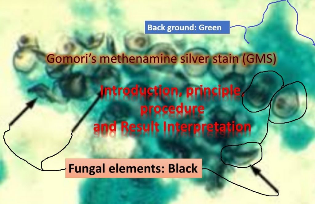
Gomori’s methenamine silver stain (GMS) is a Grocott’s modification that delineates fungal elements sharply in black against a pale green background. It is especially useful as a histopathological tool and for the detection of Pneumocystis jiroveci.
Principle of Gomori’s methenamine silver stain
The cellular walls of fungi are very thick and contain much more carbohydrates. Chromic acid creates dialdehyde groups from the carbohydrates of the fungi cell walls with over oxidation and destroys the carbohydrate structures in the tissue section. Gomori’s methenamine silver stain utilizes a light green counterstain, resulting in fungus cell walls that are various shades of black.
Procedure of Gomori’s methenamine silver stain
- Dry the smear and then fix it in absolute methanol for 5 minutes.
- Wash in distilled water.
- Dip slide in Coplin jar containing 4% chromic acid for 45
minutes. - Wash in distilled water.
- Add 1% sodium/potassium metabisulphite for 1 to 2 minutes.
- Wash in distilled water.
- Dip slide in working solution of hexamine (which is preheated in
a water bath to 56°C) for 1 hour (smear becomes dark brown). - Wash with distilled water, or if smear turns black wash with
0.1% FeCl3 - Wash with 5% sodium thiosulphate for 2 minutes.
- Wash with distilled water.
- Wash with 1% light green solution for 1 minute.
- Dry and view under oil immersion
Observation of Gomori’s methenamine silver stain
Focus the slide at 10X objective and finally observe at 100X objective i.e. oil immersion fields.
Result Interpretation of Gomori’s methenamine silver stain
- The positive control slide of Pneumocystis jiroveci
Fungal elements: black
Background: green
Cysts: dark centers, cup-shaped crescents, and cysts with fold-like lines (resembling punched-in ping pong balls)
- Fungi and actinomycetes: grey to black.
- Glycogen, mucin, and red blood cells: rose/dark grayish
brown/gray.
Disadvantages of Gomori’s methenamine silver stain
Problems with the GMS are due to non-specific staining – Proper oxidation must be used to reduce the staining of reticulum and other carbohydrate moieties that have a weaker reaction with silver than do oxidized fungal walls. Under oxidation will not generate adequate aldehyde formation to produce adequate staining. Over staining produces loss of detail and non-specific silver deposition. Under staining may lead to a misdiagnosis when only rare organisms are present e.g. Histoplasmosis – When the tissue is under stained, few organisms may not be visualized, resulting in a false-negative diagnosis
Modification in GMS and Related Stains
Churukian and Schenk have developed an oxidation- reduction silver staining method that utilizes microwave heating and can be applied to unprocessed and unfixed specimens. This method is based on a 5% periodic acid oxidizing solution followed by an ammoniacal lithium carbonate silver solution. It also can be used on deparaffinized sections, but the tissue adheres better to slides if 5% chromic acid is substituted for 5% periodic acid. It helps in the quick diagnosis of fungal and Pneumocystis jiroveci infections.
Bibliography
- Bancroft, J.D. and Stevens, A.: theory and practice of histological techniques ed.7, Churchill Livingstone inc. 1990. Edinburgh. London, Melbourne, and New York.
- C.F.A. Culling, Handbook of Histopathological and Histochemical Techniques, 1974, 671.
- Grizzle WE: Theory and practice of silver staining in histopathology. J Histotechnol 19(3):183–195, 1996.
- file:///C:/Users/Berry/Pictures/B4416.pdf