
KOH Mount of Ear Discharge Showing Septate Hyphae and Conidia of Aspergillus fumigatus
KOH mount of Ear Discharge
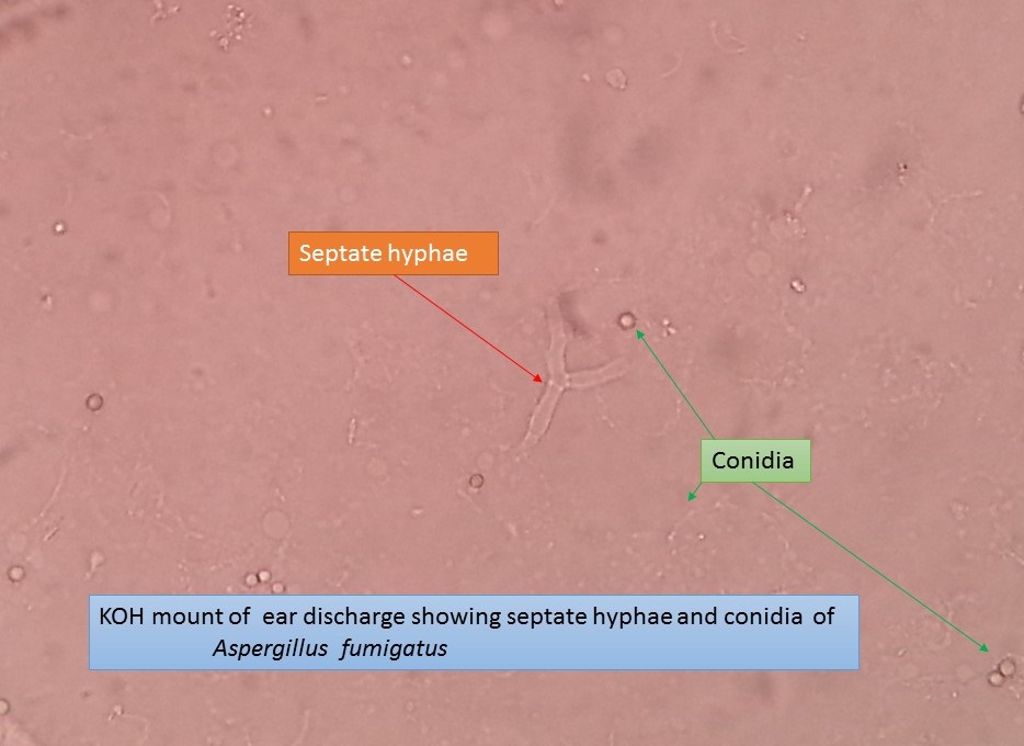
KOH mount of ear discharge showing septate hyphae and conidia of Aspergillus fumigatus as shown above picture . It conformed later culturing on SDA with the help of colony morphology and LPCB tease mount.
KOH Mount Preparation
Principle- The fungal cell wall glucans being alkali resistant, is not dissolved in KOH and other skin layer, keratin, mucus and other tissue components are dissolved.
10 or 20 or 30 % KOH is used. Skin, nail, hair or other samples obtained are mixed with a drop of KOH on a slide and covered with a cover slip. Heat may be applied to potentiate the clearing of tissue. If the specimen is transparent it may be examined without treatment with KOH. Malassezia furfur shows oval or bottle shaped cells along with short, curved hyphae. Hortae werneckishows brownish, branched, septate hyphae and budding cells. Trichosporon beigelli shows hyphae and rectangular arthrospores within(endothrix) and around(exothrix) hair. Piedraia hortae shows dark color septate hyphae around hair and asci containing 2-8 aseptate ascospores. Dermatophytes in skin scraping, nail clippings or nail scraping and hair stubs can appear as refractile, hyaline, septate, branched or unbranched hyphae and arthrospores. Several modifications of the basic 10% KOH preparation have been made for more rapid detection. This includes incorporating Parker superchrome blue in the KOH solution for selective staining of the fungus.
Other modifications of the basic KOH mount includes
- Addition of 36% of DMSO to 20% KOH to clearing of the specimen without heating.
- Addition of 5-10% glycerine to the KOH preparation to delay crystallisation of the KOH, degrading of the fungus and dehydration.
- In KOH preparation it is very difficult to differentiate ‘mosaic fungus’ (cholesterol crystals deposited around the periphery of the epidermal cells). It can be recognized by lack of internal organelles.
- Parker’s ink can also be used for better visualisation of Malassezia furfur and (Scopuloropsis spp. and Candida spp.) causing onychomycosis.
KOH with calcofluor white
Calcofluor white is a whitening agent used in the textile and paper industry. A drop of calcofluor white (a fluorescent dye) can be added to the KOH preparation before adding a cover slip. Calcofluor white binds to the polysaccharide present in the chitin or to cellulose. Fungal element fluoresce apple green or blue white. So, any element with a polysaccharide skeleton will fluoresce. Although background elements may also fluoresce, the fungal components are brighter.
Further Readings
- Rippon’s JW: Medical Microbiology. The pathogenic fungi and the pathogenic Actinomycetes. 3rd ed 1988 Publisher WB saunder co, Philadelphia.
- Clinical Microbiology Procedure Hand book Vol. I & II, Chief in editor H.D. Isenberg, Albert Einstein College of Medicine, New York, Publisher ASM (American Society for Microbiology), Washington DC.
- A Text Book of Medical Mycology. Editor: Jagdish Chandar. Publication Mehata, India.
- Practical Laboratory Mycology. Editors: Koneman E.W. and G.D. Roberts, 3rd ed 1985, Publisher Williams and Wilkins, Baltimore.
- Text book of Diagnostic Microbiology. Editors: Connie R. Mahon, Donald G. Lehman & George Manuselis, 3rd edition2007, Publisher Elsevier.
- Mackie and Mc Cartney Practical Medical Microbiology. Editors: J.G. Colle, A.G. Fraser, B.P. Marmion, A. Simmous, 4th ed, Publisher Churchill Living Stone, New York, Melborne, Sans Franscisco 1996.
- Bailey & Scott’s Diagnostic Microbiology. Editors: Bettey A. Forbes, Daniel F. Sahm & Alice S. Weissfeld, 12th ed 2007, Publisher Elsevier.