
Introduction of Microbiology Color Atlas
The name ‘ Microbiology color atlas’ is given even due to the vast spectrum of microbiology but puny collection and another thing are that only an epic centre collection of my authentical performance. So, please if you have benefited from this atlas, let others know about it too and share them through social media.

Microbiology color atlas collections
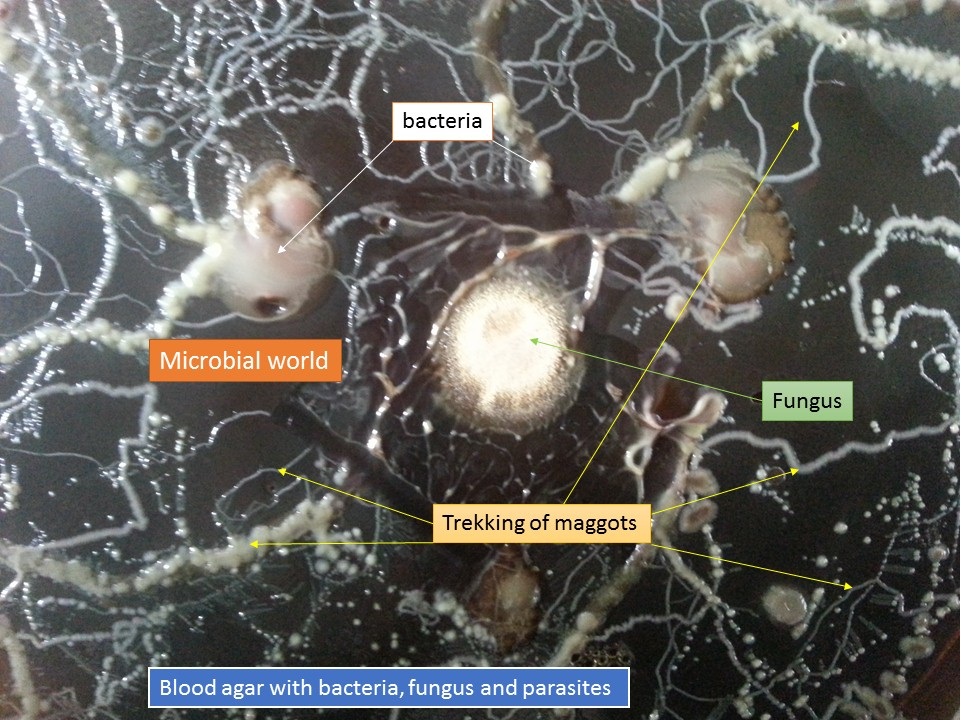
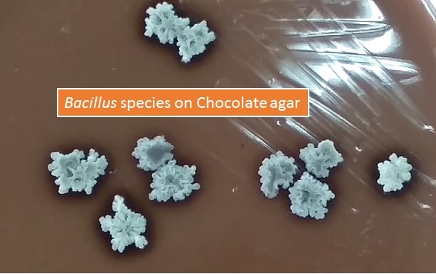
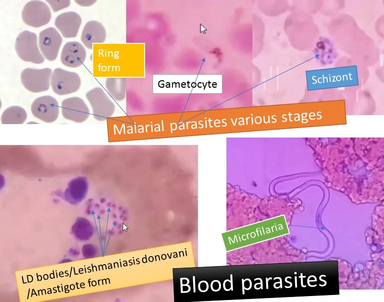
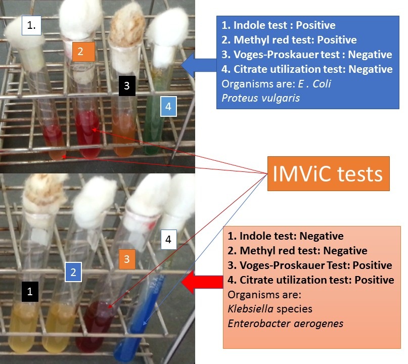
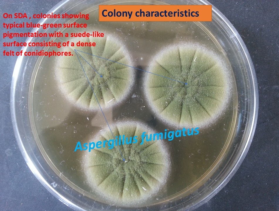
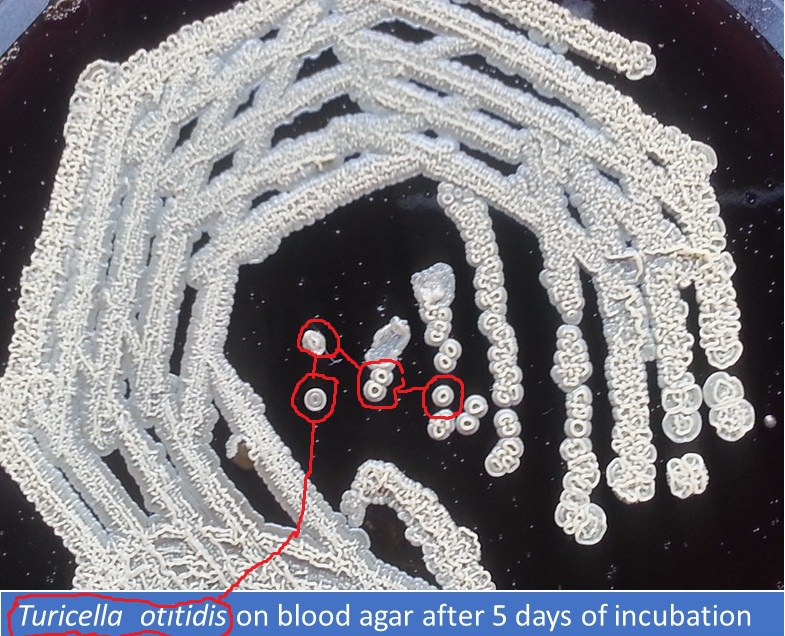
Here you will get a random collection of images pertaining to clinical/ medical microbiology i.e. bacteriology, virology, parasitology, mycology, serology/ immunology, and such related fields.


The encapsulated strain of Klebsiella pneumoniae

Clostridium spp. growth on BAP

Corynebacterium diphtheriae colonies on Blood agar

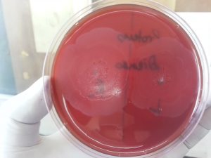
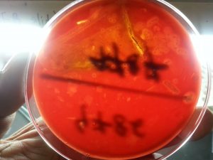
Dienes phenomenon of Proteus spp.

D-Zone test Positive Staphylococcus aureus

Kingella kingae on Gram-stained Picture lying in pairs

Mannitol Salt Agar showing yellow colonies of Staphylococcus aureus

Many Mycobacterium-tuberculosis-acid-fast-bacilli-AFB-in-Auramine-phenol-fluorochrome-stained-smear


AFB-on-Z-N-Stained-picture-with-Methylene-blue-as-counter-stain-

Clostridium-Gram-stained-picture-with-occasional-spores
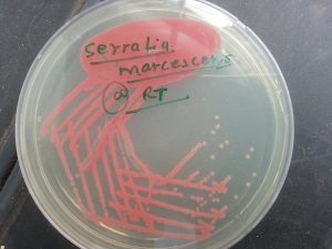
Prodigiosin-Pigment-expression of Serratia marcescens

Providencia-rettgeri-biochemical-set

Urease positive Mycobacterium tuberculosis
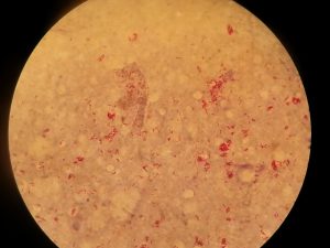
Red-bacilli-single-clump-and-globi-in-Ziehl-Nielsen-cold-stained-smear-of-skin
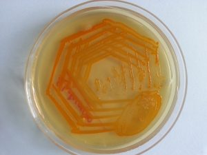
Rhodococcus-Salmon-Pink-Colonies on Nutrient agar
Beta-haemolytic-colonies-on-BAP-of-Kingella-kingae


Books Used for Identifications
- Cowan & Steel’s Manual for identification of Medical Bacteria. Editors: G.I. Barron & R.K. Felthani, 3rd ed 1993, Publisher Cambridge University Press.
- Bailey & Scott’s Diagnostic Microbiology. Editors: Betty A. Forbes, Daniel F. Sahm & Alice S. Weissfeld, 12th ed 2007, Publisher Elsevier.
- Clinical Microbiology Procedure Handbook Vol. I & II, Chief in editor H.D. Isenberg, Albert Einstein College of Medicine, New York, Publisher ASM (American Society for Microbiology), Washington DC.
- Colour Atlas and Textbook of Diagnostic Microbiology. Editors: Koneman E.W., Allen D.D., Dowell V.R. Jr, and Sommers H.M.
- Jawetz, Melnick and Adelberg’s Medical Microbiology. Editors: Geo. F. Brook, Janet S. Butel & Stephen A. Morse, 21st ed 1998, Publisher Appleton & Lance, Co Stamford Connecticut.
- Mackie and Mc Cartney Practical Medical Microbiology. Editors: J.G. Colle, A.G. Fraser, B.P. Marmion, A. Simmons, 4th ed, Publisher Churchill Living Stone, New York, Melbourne, Sans Francisco 1996.
- Textbook of Diagnostic Microbiology. Editors: Connie R. Mahon, Donald G. Lehman & George Manuselis, 3rd edition2007, Publisher Elsevier.
- Medical Mycology. Editors: Emmons and Binford, 2nd ed 1970, Publisher Lea and Febiger, Philadelphia.
- Rippon’s JW: Medical Microbiology. The pathogenic fungi and the Pathogenic Actinomycetes. 3rd ed 1988 Publisher WB Saunder co, Philadelphia.
- Clinical Microbiology Procedure Handbook Vol. I & II, Chief in editor H.D. Isenberg, Albert Einstein College of Medicine, New York, Publisher ASM (American Society for Microbiology), Washington DC.
- A Textbook of Medical Mycology. Editor: Jagdish Chander. Publication Mehata, India.
- Practical Laboratory Mycology. Editors: Koneman E.W. and G.D. Roberts, 3rd ed 1985, Publisher Williams and Wilkins, Baltimore
- Topley & Wilsons Medical Mycology. Editors: M.T. Parker & L.H. Collier, 8th ed 1990, Publisher Edward Arnold publication, London.
- Textbook of Diagnostic Microbiology. Editors: Connie R. Mahon, Donald G. Lehman & George Manuselis, 3rd edition2007, Publisher Elsevier.