
Haemophilus influenzae: Introduction, Morphology, Pathogenecity, Laboratory Diagnosis, Treatment and Key Notes
Introduction of Haemophilus influenzae
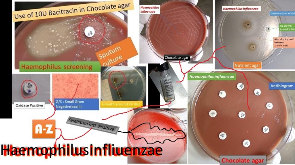
Haemophilus influenzae colonies on chocolate agar after overnight incubation in a carbon dioxide atmosphere as shown above image. It must contain haemin or other iron-containing porphyrin and nicotinamide adenine dinucleotide (NAD) or its phosphate (NADP). The porphyrin requirement is referred to as growth factor X and the NAD or NADP requirement as growth factor V. It was a causative agent of the devastating 1918 pandemic of influenza to now from local to systemic infections.
Classification of Haemophilus
- Domain: Bacteria
- Kingdom: Eubacteria
- Phylum: Proteobacteria
- Class: Gammaproteobacteria
- Order: Pasteurellales
- Family: Pasteurellaceae
- Genus: Haemophilus
- Species: H. influenzae
History of Haemophilus
- First described in 1892 by Richard Pfeiffer during an influenza pandemic
- Isolated by Smith, Andrewes, and Laidlaw in 1933
- Need one or both of accessory growth factors X and V present in the Blood
General Features of Haemophilus
- Small
- Non-motile
- Non-sporing
- Oxidase test positive
- Pleomorphic Gram-negative rods
- Transmitted via respiratory droplets, or direct contact with contaminated secretions
- Normal flora of the human respiratory tract and oral cavity
Clinical Importance of Hamophilus
- H. influenzae type b is an important human pathogen
- H. ducreyi is a sexually transmitted pathogen causing chancroid.
- Other Haemophilus are normal flora like H. parainfluenzae – pneumonia and endocarditis, H. aphrophilus causes pneumonia and endocarditis and H. aegyptius causes pink eye (purulent conjunctivitis).
Haemophilus Influenzae
- Aerobic gram-negative bacteria
- Polysaccharide capsule
- Six different serotypes (a-f) of the polysaccharide capsule
- 95% of invasive diseases caused by type b (Hib) Haemophilus Influenzae (formerly called Pfeiffer’s bacillus or Bacillus influenzae )
Morphology of Haemophilus Influenzae
It is small having a size of 1.5 x 0.3 µm, Gram-negative, non -motile rods showing considerable pleomorphism. It is non-sporing and non-acid fast. The cells are usually cocobacilliary in young cultures ( 18-24 hours), while in older cultures, long filamentous forms may be seen. Pleomorphic appear as clusters or coccobacillus forms in sputum sample while infected CSF ( meningitis) long and filamentous form predominates.
Cultural Characteristics of Haemophilus Influenzae
H. influenzae needs fastidious growth requirements. Factors X and V are essential for growth. X is hemin heat stable and porphyrins for the synthesis of cytochromes. V factor is coenzyme nicotinamide adenine dinucleotide (NAD) phosphate acts as a hydrogen acceptor. It grows well on chocolate agar and it is a non-selective, enriched growth medium that is the lysed blood agar. The name of agar is for its color when the red blood cells (RBCs) lysis gives the medium a chocolate-brown color without having chocolate products. It uses for the isolation of fastidious bacteria, such as Haemophilus influenzae, when incubated at 35-37°C in a 5% CO2 incubator. Haemophilus influenzae shows opaque cream to gray colonies.
H. influenzae in a Gram stain of a sputum sample appears as Gram-negative coccobacilli. Haemophilus influenzae requires X and V factors for growth and therefore, it grows only around the paper disk that has been impregnated with X and V factors on non-selective and non-enriched media like nutrient agar or MHA. There is no bacterial growth around the disk that only contains either X or V factor but may be in between X and V disks as shown above picture. It also shows satellitism. When Staphylococcus aureus is streaked across a plate of blood agar with a species containing H. Influenzae the colonies which are large development along with the streak of Staphylococcus and are small further away as shown above image and below video.
Biochemical Reactions of Haemophilus Influenzae
H. influenzae is catalase and oxidase test positive ferments glucose and galactose, reduces nitrate to nitrite. It does fement lactose, mannitol and sucrose. On the basis of production of urease, indole, and ornithine decarboxylase, it is divided into 8 biotypes.
Biotypes Urease Indole Ornithine decarboxylase
- I + + +
- II + + –
- III + – –
- IV + – +
- V – + +
- VI – – +
- VII – – –
- VIII – – –
Resistance
It is a delicate organism and it is readily killed by heating at 55°C for 30 minutes. Refrigeration, drying, and disinfectants destroy the organisms. For long-term preservation, the culture may be lyophilized.
Antigenic Structures of Haemophilus Influenzae
It contains 3 major surface antigens and they are-
- Capsular polysaccharide
- Outer membrane proteins (OMP)
- Lipopolysaccharides ( LPS )
Pitman Classification– The major antigenic determinant of capsulated strains into six capsular types type a to f. Typing by agglutination reaction using type-specific antisera. Other methods include-
- Quelling reactions
- Coagglutination
- Countercurrent electrophoresis ( CIEP)
- ELISA
Type b characteristics
Hib has unique characters contains pentose sugars, ribose ribitol, instead of hexose in others, and hexosamines. The capsular polyribosyl ribitol phosphate ( PRP ) of Hib induces IgG, IgM, and IgA antibodies which are bactericidal, opsonic, and protective. So Hib PRP ( polyribosyl ribitol phosphate) was employed for Immunization.
Mode of Transmission
Droplet infection and discharge from the upper respiratory tract (URT) during the infectious period. The incubation period is unknown, probably short, 2-4 days. Infectious Period – As long as the organism is present, even in the absence of nasal discharge. Non-infectious within 24 to 48 hours after the start of effective antibiotics.
Haemophilus influenzae type b Clinical Features
- Epiglottitis
- Meningitis
- Pneumonia
- Osteomyelitis
- Arthritis
- Cellulitis
- Bacteremia
Non -typable Strains
H. Influenzae lacking capsules that are nontypable are most relevant in clinical infections. Outer membrane proteins (OMP) of Hib are classified into 13 subtypes. H. Influenzae lipopolysaccharides are more complex. The genome of the organism is sequenced.
Pathogenicity
A human pathogen can produce invasive and non-invasive lesions. The prominent organism is producing meningitis. It can produce laryngoepiglottitis, conjunctivitis, bacteremia, pneumonia, arthritis, endocarditis and pericarditis. Most strains are opportunistic pathogens. Most strains of H. influenzae are opportunistic pathogens; that is, they usually live in their host without causing disease, but cause problems only when other factors (such as a viral infection or reduced immune function) create an opportunity.
Most Important Clinical Illness associated with H. influenzae
Pneumonia: Severe shortness of breath, rapid heart rate, fever, cough, and evidence of pneumonia by chest radiograph.
Septic Arthritis: Swelling, warmth, pain with movement and decreased mobility of a single large weight-bearing joint.
Haemophilus Influenza
Mode of Transmission
Droplet infection and discharge from the upper respiratory tract during the infectious period. Incubation Period Unknown, probably short, 2-4 days. The infectious period is as long as the organism is present, even in the absence of nasal discharge. Non-infectious within 24 to 48 hours after the start of effective antibiotics.
Haemophilus influenzae type b meningitis
Accounted for approximately 50%-65% of cases in the prevaccine era. Hearing impairment or neurologic sequelae in 15%-30%. The case-fatality rate is 2%-5% despite effective antimicrobial therapy.
Secondary Infections: Respiratory tract infections, otitis media, Sinusitis, chronic bronchitis
Haemophilus Meningitis: It carries high mortality of 90% if not treated. The Bacteria reach meninges from the nasopharynx.
Laryngol epiglottitis: Causes epiglottis, obstructive laryngitis, > 2 years children are vulnerable which may be fatal in 2 hours.
Pneumonia: Pneumonia along with Meningitis, Lobar Pneumonia, bronchopneumonia. It can present with empyema ( a collection of pus).
Suppurative Lesions: Arthritis, endocarditis, pericarditis, hematogenous dissemination, otitis media, cellulitis
Laboratory Diagnosis of Haemophilus Influenzae
Specimens: It depends on the site of infection, the following specimens may be collected-
Collection and Transport of specimens
Specimens should be collected in sterile conditions and under all aseptic conditions.
Microscopy
Gram stain: Gram-stained smear of CSF in meningitis shows pleomorphic long and filamentous form predominate while in sputum coccobacillary forms.
Immunofluorescence and quellung reaction: For a direct demonstration of H. influenzae after mixing with specific tube b antiserum.
Antigen detection: Type b capsular antigen can also be detected in patient serum, CSF, urine, or pus by following methods-
- Latex agglutination
- Coagglutination
- Counter immunoelectrophoresis (CIE)
Culture
CSF culture- Inoculate CSF on chocolate age and incubate the plate at 35-37 °C, aerobically with 5-10 % CO2, overnight. After that, identify on the basis of colony morphology, gram staining, oxidase test, satellitism, serotyping, etc.
Blood culture: It is useful in cases of pneumonia and epiglottitis.
Sputum culture
Colony Morphology and Staining
Serotyping: It may be performed with type-specific antisera as shown above picture.
Treatment
Following antimicrobial drugs are useful to treat and they are-
- Cefotaxime
- Ceftazidime
- Ampicillin
- Contrimixazole
- Amoxycillin with Clavulanate
- Clarithromycin
Epidemiology and Prevention
- Similar to Pneumococci
- The infection enters through the respiratory tract
- Immunity is type-specific
- HIB is protected by the PRP vaccine
- Poorly immunogenic in children below 2 years
- Rifampicin can be given for 4 days and prevents secondary infection and eradicates carrier state.
Public Health Action
Current Vaccines: Haemophilus B conjugate vaccine is widely using Haemophilus influenzae type b vaccine has reduced H. influenzae type b meningitis in children by 95%.
Newer vaccines: The previous vaccines’ PRP is immunogenic in older children. PRP is poorly immunogenic in children below two years and Immunogenicity can be improved when coupled with protein carriers like diphtheria and tetanus toxoid that should be used in young children.
Key Notes on Haemophilus
- Virulent strains possess capsules while the avirulent strains and older cultures are non-capsulated.
- 95% of H. Influenzae isolates belong to type b in the Pitman classification.
- Plasmid born resistance set in Ampicillin
- H. ducreyi a causative agent of Chancroid in bipolar stain shows bacilli in small groups appear as parallel chains giving school of fish appearance.
- Haemophilus aegypticus is also called Koch – Weeks Bacillus. Identified as Biotype of H. influenzae that produces pink eye infection for which sulphonamides and gentamycin are effective antimicrobial agents.
- H. aphrophilus requires X and V factors that produce bacterial endocarditis, brain abscess, sinusitis, and abscess.
- H. influenzae biogroup aegyptius causes Brazilian Purpuric Fever.
- Haemophilus influenzae type b medical management requires hospitalization and treatment with an effective 3rd generation cephalosporin, or chloramphenicol plus ampicillin.
- Ampicillin-resistant strains of HIB are now common throughout the United States.
- As you know, H. influenzae is very sensitive to low temperature and thus, clinical samples should never be refrigerated, instead, samples should be transported to the laboratory without delay and inoculated on culture media immediately.
Related Text and Video
Various Haemophilus species Identification using X, V, and XV test
Haemophilus spp. have varying requirements for X, V, and XV growth factors. Consequently, the significant differences in growth factor requirements of Haemophilus spp. allows for their differentiation. Differentiation is based on the presence or absence of growth around and/or between discs impregnated with factors X, V, and XV.
Each X-Factor Disk is impregnated with hemin. Each V-Factor Disc is impregnated with NAD (nicotinamide adenine dinucleotide). Each XV-Factor Disc is impregnated with a combination of hemin and NAD.

#Variety of Haemophilus species identification on basis of X, V, and XV disks, blood agar, and Xylose test as shown below-
Haemophilus influenzae on chocolate agar
Haemophilus influenzae colonies on chocolate agar after overnight incubation in a carbon dioxide atmosphere as shown below the video. It must contain haemin or other iron-containing porphyrin and nicotinamide adenine dinucleotide (NAD) or its phosphate (NADP). The porphyrin requirement is referred to as growth factor X and the NAD or NADP requirement is growth factor V.
Tool and techniques for isolation of Haemophilus influenzae as shown below-
Introduction of Chocolate agar
Chocolate Agar (CHOC) is a non-selective, enriched growth medium that is the lysed blood agar. The agar is named for its color when the red blood cells (RBCs) lysis gives the medium a chocolate-brown color without having chocolate products. It is used for the isolation of fastidious bacteria, such as Haemophilus influenzae, when incubated at 35-37°C in a 5% CO2 incubator.
Principle of Chocolate Agar
The composition of chocolate agar is the same as the blood agar and the only difference is while preparing Chocolate agar, the red blood cells are lysed changing the medium color chocolate brown.
The lysis of RBC during the heating process releases intracellular coenzyme nicotinamide adenine dinucleotide (Factor V or NAD) into the agar for utilization by fastidious bacteria (the heating process also inactivates growth inhibitors). Hemin (factor X) is available from non-hemolyzed as well as hemolyzed blood cells.
The most common species that require this enriched medium for growth include Neisseria meningitidis and Haemophilus spp. H. influenzae is not able to grow on sheep blood agar.
Requirements for Chocolate Agar Preparation
- Prepared blood agar
- Incubator for the simplest method
- But for other methods are
- Blood agar base
- Sheep blood
- Distilled water
- Measuring cylinder
- Autoclave
- Weighing balance
- Water bath
- Hot air oven ( optional)
Preparation of Chocolate Agar
It can be prepared by the following methods.
Simplest method
Take already prepared blood agar plates (5% sheep blood agar) and put those plates into a hot air oven for 2 hours at 55°C.
Take out those plates and you will get chocolate agar.
Place the plates in sterile plastic bags and store them at 4°C until use.
As a sterility test, incubate an uninoculated plate for 48 hours at 35-37°C with 5% CO2.
Another Method
- Suspend 40.5 grams in 1000 ml distilled water or deionized water.
- Heat to boiling to dissolve the medium completely.
- Sterilize by autoclaving at 15 lbs. pressure (121°C) for 15 minutes.
- Heat-lyse a volume of sheep blood that is 5% of the total volume of media being prepared very slowly to 56°C in a water bath.
- Dispense 20 ml into 15×100 mm Petri dishes. Allow the media to solidify and condensation to dry.
- Place the plates in sterile plastic bags and store them at 4ºC until use.
- As a sterility test, incubate an uninoculated plate for 48 hours at 35-37°C with 5% CO2 (or in a candle-jar).
Quality Control
For the quality control inoculate N. meningitidis, S. pneumoniae, and H. influenzae QC strains inoculate into prepared chocolate agar (CHOC) for 18-24 hours at 35-37°C with 5%CO2 (or in a candle jar but it can only provide up to 3% CO2).
Organisms growth
N. meningitidis luxuriant
S. pneumoniae luxuriant
H. influenzae luxuriant
Colony Characteristics in Chocolate Agar
- Haemophilus influenzae: Non-hemolytic, opaque cream to gray colonies.
- Neisseria meningitidis: Growth on chocolate agar is grayish, non-hemolytic, round, convex, smooth, moist, glistening colonies with a clearly defined edge.
- Neisseria gonorrhoeae: Colonies on CHOC are pinkish-brown and translucent, exhibit smooth consistency and defined margins, and are typically 0.5-1 mm in diameter.
Uses of Chocolate Agar
- It is a very useful medium to isolate fastidious organisms in Microbiology Laboratory from various clinical specimens like sputum ( H. influenzae), urethral discharge ( N. gonorrhoeae), CSF/blood (N. menigitidis) .
- And thus, Chocolate agar uses to isolate and cultivate fastidious microorganisms such as Haemophilus species and Neisseria species.
- It is also useful in isolating N. gonorrheae from both acute and chronic cases of gonococcal infections.
- It is also useful in isolating N. meningitidis from bacterial meningitis.
- Chocolate agar with bacitracin acts as a selective medium for screening H. influenzae from specimens e.g. sputum containing a mixed flora of microorganisms.
- Its modified forms uses given below.
Modification of Chocolate Agar (CHOC)
- Thayer-Martin agar/medium uses for the selective isolation of N. gonorrhoeae and N. meningitidis. This Media is a chocolate agar supplemented with vancomycin, colistin, and nystatin (VCN) to inhibit the normal flora, including non-pathogenic Neisseria from the clinical specimens
- Chocolate Agar with bacitracin: CHOC with bacitracin is a selective medium uses to improve the primary isolation of Haemophilus influenzae from specimens containing a mixed flora of microorganisms.
- Chocolate agar with GC base and growth supplement: It is a medium that supports the special growth requirements (hemin and NAD) needed for the isolation of fastidious organisms, such as H. influenzae, when incubated at 35-37°C in a 5% CO2 atmosphere.
- Chocolate agar with TSA and growth supplements: It is a medium that supports the special growth requirements (hemin and NAD) needed for the isolation of fastidious organisms, such as H. influenzae, when incubated at 35-37°C in a 5%CO2atmosphere.
Culture media the simplest way of identification | Blood |MacConkey | Chocolate |RCM| Nutrient agar as shown below-
E. coli on MacConkey agar, blood agar, and Chocolate agar as shown below-
Note: There is no need of using chocolate agar for cultivation of E. coli because it can grow even on an ordinary mediums like nutrient agar, but want to share one thing is that all the organisms growing on nutrient agar/ MacConkey agar/ blood agar can easily grow on chocolate agar but not vice versa.
Campylobacter on chocolate agar-
Satellitism test Positive of Haemophilus influenzae is shown below-
Haemophilus influenzae on Gram’s stained smear under the microscope showing gram-negative cocobacilli , small to the large rod, and pleomorphic forms as shown below-
Further Reading
- Bailey & Scott’s Diagnostic Microbiology. Editors: Bettey A. Forbes, Daniel F. Sahm & Alice S. Weissfeld, 12th ed 2007, Publisher Elsevier.
- Colour Atlas and Textbook of Diagnostic Microbiology. Editors: Koneman E.W., Allen D.D., Dowell V.R. Jr, and Sommers H.M.
- Jawetz, Melnick and Adelberg’s Medical Microbiology. Editors: Geo. F. Brook, Janet S. Butel & Stephen A. Morse, 21st ed 1998, Publisher Appleton & Lance, Co Stamford Connecticut.
- https://www.thermofisher. com/order/catalog/product/R01293#/R01293
- https://www.sciencedirect.com/topics/immunology-and-microbiology/chocolate-agar
- https://www.ncbi.nlm.nih.gov/pmc/articles/PMC271442/
- https://www.slideshare.net/doctorrao/hinfluenza
- https://jcm.asm.org/content/jcm/20/4/822.full.pdf
- https://www.ncbi.nlm.nih.gov/pmc/articles/PMC533390/
- https://anaerobesystems.com/products/plated-media/chocolate-agar-choc
- Mackie and Mc Cartney Practical Medical Microbiology. Editors: J.G. Colle, A.G. Fraser, B.P. Marmion, A. Simmous, 4th ed, Publisher Churchill Living Stone, New York, Melborne, Sans Franscisco 1996.
- Manual of Clinical Microbiology. Editors: P.R. Murray, E. J. Baron, M. A. Pfaller, F. C. Tenover and R. H. Yolken, 7th ed 2005, Publisher ASM, USA
- Textbook of Diagnostic Microbiology. Editors: Connie R. Mahon, Donald G. Lehman & George Manuselis, 3rd edition2007, Publisher Elsevier.
- A textbook of Microbiology, 2nd ed, Prof. C.P. Baveja
- https://www.ncbi.nlm.nih.gov/pmc/articles/PMC4042752/
- https://www.webmd.com/children/meningococcal-meningitis-symptoms-causes-treatments-and-vaccines