
Introduction
The Dalmau plate culture is a classic mycological technique used to identify and differentiate yeast species, particularly those in the genus Candida, based on their unique microscopic morphological features.
Purpose and Mechanism
The primary goal of this technique is to induce the formation of specific fungal structures—such as pseudohyphae, blastoconidia, and chlamydospores—that do not typically appear on standard nutrient-rich media like Sabouraud Dextrose Agar. It utilizes “starvation” media that provide limited nutrients to trigger these diagnostic morphological changes.
Common Media Used
- Cornmeal Agar (CMA): Often supplemented with Tween 80 (a surfactant) to further stimulate the production of chlamydospores and pseudohyphae.
- Rice Extract Agar: Sometimes used as an alternative to CMA for similar results.
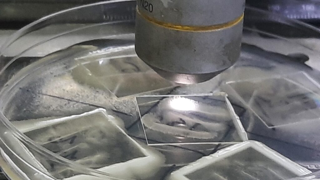
Procedure
Prepare Cornmeal agar containing 1% Tween 80 in a 90-mm plate. Divide the plate into 4 quadrants and label each quadrant. Using a sterile needle or straight wire, lightly touch the yeast colony and then make 2-3 streaks of approximately 3.5 – 4 cm long and 1.2 cm apart. Place a flame-sterilized and cooled 22 mm square cover glass over the control part of the streak. This will provide a partially anaerobic environment at the margins of the cover slip. Incubate the plates at 25°C for 3-5 days. Remove the lid of the petri plate and place the plate on the microscope stage, and observe the edge of the cover glass using the low-power objective (10X) first and then the high-power objective (40X). Morphological features like hyphae, pseudohyphae, blastospores, ascospores, chlamydospores, basidiospores, or sporangia are noted.
Key Morphological Identifiers
| Structure | Significance | Example Species |
| Chlamydospores | Large, thick-walled, round terminal cells. | Candida albicans (Classic diagnostic feature) |
| Pseudohyphae | Chains of elongated yeast cells that remain attached. | Candida tropicalis, C. parapsilosis |
| Blastoconidia | Small, budding yeast cells. | Found in most yeast species |
Note: While largely replaced in high-resource settings by automated systems like MALDI-TOF MS, the Dalmau technique remains a valuable, cost-effective method for preliminary identification in clinical laboratories.
Further Readings
- https://www.microdigest.net/2025/09/dalmau-plate-technique-cherished-in.html
- https://mycology.adelaide.edu.au/fungal-descriptions-and-antifungal-susceptibility/yeast-like-fungi
- https://www.afwgonline.com/wp-content/uploads/2018/12/1115_TanAL_Identification-of-Yeasts-lecture_Final.pdf
- https://mycology.adelaide.edu.au/candida
- https://www.remedypublications.com/open-access/identification-of-candida-species-conventional-methods-in-the-era-of-molecular-diagnosis-775.pdf
- https://www.sciencedirect.com/topics/pharmacology-toxicology-and-pharmaceutical-science/chlamydospore
- https://www.sciencedirect.com/science/article/pii/S0732889323001281
- https://mycology.adelaide.edu.au/fungal-descriptions-and-antifungal-susceptibility/yeast-like-fungi/cryptococcus
- https://www.researchgate.net/figure/The-characteristic-Dalmau-morphology-in-different-Candida-spp_tbl1_342505552